


World’s First: AI Assists Surgeons in Brain Surgery — In the Most Dangerous Confined Space at the Skull Base, Artificial Intelligence Has Stepped onto the Operating Table for the First Time as a “Navigator” to Help Surgeons Perform a High‑Difficulty Brain Surgery. This Landmark Surgery Marks AI’s Role in the Operating Room Formally Shifting from “Preoperative Consultant” to “Intraoperative Assistant.”
The world’s first AI‑assisted real‑time brain surgery was completed in the United Kingdom. On August 27, local time, University College London Hospital (UCLH) announced that during a pituitary tumor resection, AI assisted surgeons for the first time by reading endoscopic images in real time, identifying hidden blood vessels and nerves, and delineating safe resection zones, enabling precise removal of the patient’s pituitary tumor.
The pituitary gland is deeply buried at the skull base, about the size of a marble, yet surrounded by the internal carotid artery and the optic nerve. In this confined space, a surgical scalpel deviation of even 1 millimeter could mean blindness, stroke, or even death. With AI’s real‑time assistance, the surgery was a complete success. The patient has now fully recovered, with significantly improved vision.
The innovation of this surgery lies in the fact that, in the past, AI in the operating room was primarily responsible for preoperative image reading, path planning, or postoperative review, quality control, and teaching. Now, for the first time, it has participated in the surgical process in real time.
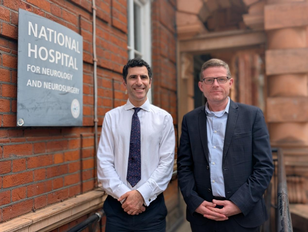 Fig.1.A photo of the lead surgeons Hani Marcus (left) and Rhys Hibbert (right)
Source: UCLH
A Surgery That Was Postponed
The patient who underwent this pioneering surgery was Rhys Hibbert, a 48‑year‑old British man who works as a customer service manager and volunteers as an ambulance worker.
The incident began with an unprovoked fainting episode. In December 2024, after lunch, Rhys went for a walk as he had done for years. While walking, he suddenly felt unwell, lost consciousness, and fell to the ground with convulsions. It was the first seizure of his life.
A brain scan after hospitalization revealed a tumor about 11 mm in diameter, roughly the size of a soybean, growing on the pituitary gland, which is normally only the size of a marble. The pituitary gland is located at the center of the brain, directly behind the root of the nose — hidden but critically important. It is the “command center” of the body’s endocrine system, responsible for secreting hormones that regulate growth, metabolism, blood pressure, and body temperature.
Initially, doctors recommended conservative observation. But the tumor grew faster than expected, and the symptoms of hormonal imbalance became increasingly pronounced. Rhys experienced extreme fatigue, dizziness, and his visual field progressively narrowed — like a piece of paper covering the lower half of a screen. He began tripping over things frequently and had to use a cane.
The psychological impact was even greater. “There were many kind people around me, but I didn’t want to be a burden to anyone. I’ve been married for 18 years, and every morning I make a cup of coffee for my wife — I didn’t want the tumor to change that,” Rhys said. Ultimately, he decided to undergo endoscopic endonasal transsphenoidal pituitary tumor resection.
In this procedure, a thin endoscope with a high‑definition camera at its tip is inserted through the nostril, navigating along the gap between the middle turbinate and the nasal septum to reach the sphenoid sinus opening. The surgeon then layers through the sphenoid sinus cavity, the sella turcica floor, and the dura mater to finally remove the tumor.
This is one of the most precision‑demanding procedures in neurosurgery. On one hand, the sphenoid sinus is the “safe entry” for the surgery, but each patient’s anatomy is different — a slight error could mean cutting in the wrong place. On the other hand, the path from the sphenoid sinus to the pituitary is lined with critical nerves and blood vessels, adjacent to the internal carotid artery that supplies blood to the brain, and just above is the optic nerve that controls vision. “In this area, a scalpel deviation of just 1 millimeter can have catastrophic consequences,” the UCLH press release stated.
“The most serious risk in pituitary surgery is injury to the artery. If an accident occurs, the only emergency measure is to occlude the artery, which would lead to stroke and death,” explained Hani Marcus, a consultant at the National Hospital for Neurology and Neurosurgery (NHNN) and Professor of Neurosurgery at the UCL Queen Square Institute of Neurology.
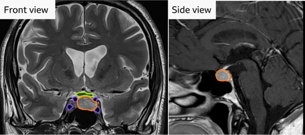 Fig.2.Frontal (left) and lateral (right) views of the brain scan showing the tumor adjacent to critical blood vessels
Source: UCLH
A few days before the surgery, Hani asked Rhys whether he was willing to become the first person in the world to undergo brain surgery with real‑time AI assistance. Rhys agreed without hesitation.
On the day of the surgery, he also brought two boxes of chocolates for the medical team. “A surgery requires the joint effort of dozens of people — it’s the work of the surgical, anesthesia, nursing, and entire support teams. I want to thank every one of them,” he said.
AI Finds the Hidden Vessels and Nerves
Above Rhys’s operating table were two screens, one of which was dedicated to displaying AI information. During the surgery, AI read the endoscopic images in real time, rapidly marking critical blood vessels, nerves, and the relatively safe resection zone.
If this surgery were compared to an off‑road rally, AI was the “navigator”in the passenger seat, responsible for guiding the surgeon safely through every turn.
The system was developed by the UCL Hawkes Institute and runs on NVIDIA’s Clara IGX platform, which is designed specifically for real‑time AI in medical devices. The clinical trial was funded by the UK’s National Institute for Health and Care Research (NIHR) and other organizations.
Sophia Bano, Associate Professor of Robotics and Artificial Intelligence at the UCL Department of Computer Science and the technical lead for the project, explained: “The system was trained on hundreds of surgical videos. We asked neurosurgeons to manually outline the contours of the internal carotid artery and other vessels and nerves frame by frame, and then fed these ‘correct answers’ to the AI.”
So, does AI “navigation” actually work? In November 2024, the research team published the answer in npj Digital Medicine. The results showed that with AI assistance, surgeons’ accuracy in identifying the sella turcica floor — the “safe entry zone” — improved from 70.7% to 77.5% (DICE coefficient), a statistically significant difference. In the critical task of pinpointing the center of the safe zone, nearly all participants hit the bullseye after AI assistance.
The greatest beneficiaries were junior surgeons. With AI assistance, medical students’ recognition accuracy jumped from 66.2% to 78.9%, matching or even surpassing that of some experienced surgeons.
In addition, AI significantly reduced the most dangerous “false positive” judgments, preventing surgeons from mistakenly identifying the internal carotid artery region as a safe zone for incision.
The study also found that different surgeons manually outlined different ranges for the same image — precisely the kind of “localization” problem that AI excels at solving. After extensive training, the system’s localization accuracy for critical structures improved substantially, and its inference speed reached real‑time levels.
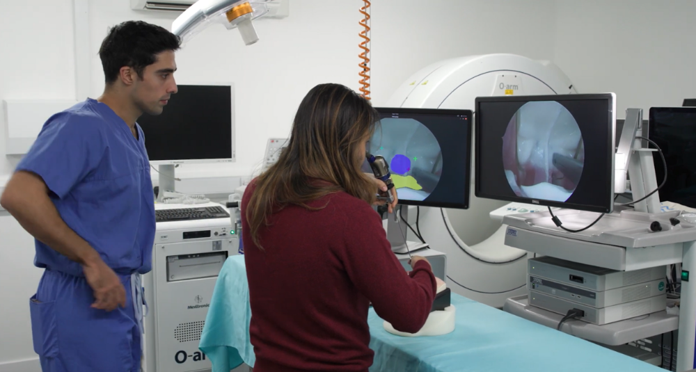 Fig.3.An image showing the AI‑navigated endoscopic endonasal transsphenoidal surgery being tested by Hani’s team. The purple area on the left screen indicates the AI‑annotated safe surgical zone; the right screen shows the nasal endoscopic view
Source: UCL Hawkes Institute
The Scalpel Remains in the Surgeon’s Hand
With AI assistance, Hani’s team successfully removed Rhys’s pituitary tumor.
“When I woke up after surgery and opened my eyes, I could see everything in the room clearly. It felt like I had a 360‑degree panoramic view — I hadn’t seen this clearly in over a year!” Rhys recalled. One week after surgery, he was walking independently without a cane, and his preoperative fatigue and other symptoms had resolved, with continued improvement in his vision.
The surgery has drawn widespread attention, and the most common question is: when will it benefit more specialties and more patients?
“The answer is: not anytime soon,” Hani candidly admitted. He explained that this surgery was performed within the framework of a clinical trial — it was the “first human application”of the AI system. It proved that the technology can work in a real operating room, but it does not yet answer questions such as “how much does AI improve the gross total resection rate?” or “how much does it reduce complications?” The team is now preparing for a larger clinical trial. “Our goal is far more than just giving surgeons an extra pair of eyes. In the future, this system will also learn to track the position of surgical instruments in real time, identify interactions between instruments and critical tissues, and provide more feedback to the surgeon.”
In fact, Chinese teams are also active on the track of AI‑assisted surgery and diagnostics.
The team led by Prof. Yugang Hong at Renmin Hospital of Wuhan University has developed ENDOANGEL, which can read endoscopic images in real time and provide immediate prompts to the physician. In July 2021, The Lancet Gastroenterology & Hepatology published the team’s prospective, single‑center, randomized controlled study, confirming that ENDOANGEL can reduce the miss rate of gastric tumors and unnecessary biopsies without prolonging examination time.
The team of Fan Zhang and Haisheng He at Fudan University, in collaboration with the teams of Lunxiu Qin and Wenwei Zhu at Huashan Hospital, Fudan University, developed a tissue autofluorescence near‑infrared imaging (TANI) technique that does not require injection of contrast agents. Published in Nature Biomedical Engineering, it can delineate the boundaries of hepatocellular carcinoma and metastatic liver tumors in real time during actual liver resection surgery, “illuminating” tumors that are otherwise invisible to the naked eye.
In Sophia’s view, the value of AI‑assisted surgery extends far beyond reducing the risk of a single operation. “AI can distill the decades of experience accumulated by top surgeons into a reproducible, distributable system, thereby bridging the most difficult‑to‑close gap in competence between physicians of different regions and different levels of seniority.”
“During surgery, AI is the surgeon’s assistant, not the decision‑maker. The scalpel remains in the surgeon’s hand,”Hani emphasized.

