


A recent case report in Nature Communications described a rare pair of identical twins in whom a malignancy crossed in utero from one fetus to the other through the shared placenta. Genetic tracing reconstructed the transplacental transmission event that occurred before birth.
This is the rare experience of identical twin sisters Amelia and Amaya. During prenatal examination, a mass was identified on the face of one fetus. When the two were delivered early by cesarean section, both were found to have tumor foci and both ultimately died.
Whether the same disease arose independently in both, or whether one twin’s tumor spread to the other, was unclear until a study published on Oct 6 in Nature Communications revealed the answer: the tumor originated in one fetus and then spread to the other in utero.
Researchers at the Wellcome Sanger Institute and Great Ormond Street Hospital, UK, analyzed 45 samples from the two infants and the placenta. The subtle somatic variants accumulated during cell division not only pointed to the tumor’s origin but also gave the prenatal disease a traceable timeline.
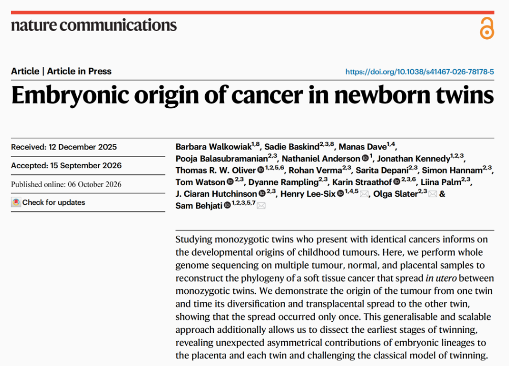 Fig.1. Article Screenshot
Article Key Points
- Nature Communications case report (Oct 6, 2026): identical twins Amelia and Amaya with in utero tumor transmission
- Whole-genome sequencing of 45 samples (tumor, normal, placenta) traced the tumor to twin A
- Twin B’s tumor cells carried A-lineage somatic variants, confirming common origin
- Driver MN1-ZNF341 gene fusion present in all tumor samples from both infants
- Mutation accumulation timing: driver fusion in late first trimester; transmission in late second trimester
- Twin A’s spleen sample showed ~75% of cells carried twin B’s lineage markers
- Findings also challenge the classical equal-contribution model of monozygotic twinning (single case)
- Evidence status: single case report; tumor was identified as undifferentiated sarcoma (small round blue cell morphology)
Both Twins Had Tumor Foci at Birth
According to the Nature Communications paper, Amelia and Amaya were monochorionic diamniotic identical twins; each had her own amniotic sac, but they shared a single placenta.
The early prenatal course was uneventful until 33 weeks of gestation, when fetal magnetic resonance imaging identified a mass on the face of fetus A. At 34 weeks, both infants were delivered by cesarean section.
However, the tumor was no longer confined to fetus A’s face; lesions were present in multiple organs, including the brain and liver. Fetus B was found to have skin nodules at birth, with subsequent involvement of the leptomeninges; intracranial hemorrhage occurred at just 8 weeks of life.
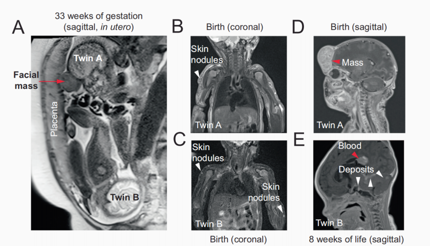 Fig.2. Clinical Imaging of the Twins
Screenshot from the Original Paper
Both infants died shortly after birth. With the family’s consent, their remains were donated for research, allowing scientists to obtain tumor, normal-tissue, and placenta samples from both, and to trace the disease to its origin.
To determine why both developed the same disease, the team first had to identify what the tumor actually was.
Skin-lesion sections from both sisters looked almost identical, with poorly defined subcutaneous nodules, dense small round blue cells showing active mitoses, but without morphological features pointing to a specific differentiation lineage. Many pediatric malignancies can present with this small round cell morphology, so morphology alone was not diagnostic.
The pathology team combined immunohistochemistry to search for additional clues. The tumor cells strongly expressed CD56, but this single marker is not sufficient to confirm neuroblastoma. PHOX2B and myogenic markers did not support neuroblastoma or rhabdomyosarcoma. Integrating morphology and immunophenotype, both tumors were most consistent with undifferentiated sarcoma.
The more decisive commonality came from molecular testing. All tumor samples analyzed from both sisters carried an MN1-ZNF341 gene fusion, which the researchers validated by RNA sequencing.
Consistent pathological features plus identical driver fusion strongly suggested a close relationship between the two tumors. But for identical twins, this still did not answer the central question: did two independent tumors arise, or did they share a common origin?
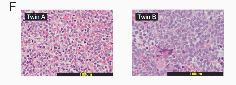 Fig.3. Small Round Blue Cell Morphology in Skin Tumor Sections of both Fetuses
Screenshot from the Original Paper
Identical twins arise from a single fertilized egg, so their genomes are highly similar, but every cell is not always perfectly identical. As the embryo develops, cells continuously divide and accumulate a small number of new somatic variants. A variant arising in one cell can be inherited by its descendant cells. Comparing which tissues carry which variants makes it possible to distinguish cell lineages and trace them back to a common ancestor.
This is the core logic of the study. The team performed whole-genome sequencing on the two infants and the placenta, including tumor and normal tissues, and used laser-capture microdissection to enrich placental trophoblast cells for sequencing.
Reconstructing the “In Utero Transplacental Transmission”
Developmental tags in normal tissue helped the researchers distinguish early cell lineages that would give rise to fetus A versus fetus B.
The results showed that all tumors, whether sampled from fetus A or fetus B, carried developmental variants of A-lineage origin. The tumor cells in fetus B retained the genetic signature of A-lineage cells.
This confirmed a common origin. The tumor first arose in fetus A and was later transferred to fetus B. Integrating clinical information with the genetic findings, the researchers concluded that the facial mass identified in fetus A was most likely the primary site.
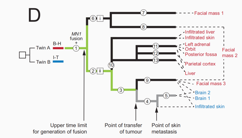 Fig.4. Clonal Lineages and Inferred Timeline of Tumor Transmission between the Twins
Screenshot from the Original Paper
Once common origin was established, the next questions were: when did the transmission occur, and by what route?
The team noted a key difference. The tumor in fetus A had already differentiated into multiple branches, with different branches seeding metastases in different organs, indicating that the cancer had been developing in A for a long time and from a complex source. Fetus B’s situation was different: her skin and intracranial tumors contained only a portion of A’s tumor branches, and these were closely related to each other.
If the two had arisen independently, fetus B’s tumor should not show a pattern of “carving out a slice of A’s tumor.” The most parsimonious explanation is that a relatively late in utero transmission event carried an entire batch of cancer cells into fetus B.
Further analysis showed that fetus B’s tumor contained at least two distinct cell branches, suggesting that more than one tumor cell was involved in this transfer event.
The number of variants also provided a temporal clue. Mutations accumulate at a roughly steady rate during cell division; the team estimated that the driver fusion emerged in the late first trimester, and transmission to fetus B occurred in the late second trimester. In other words, by the time the facial mass was detected on imaging at 33 weeks, the cancer cells had likely already entered the other fetus, though no symptoms had yet appeared.
But how did the cancer cells cross the boundary between the two fetuses?
The paper explained that the twins shared a placenta with interconnected vasculature, allowing blood and cells to be exchanged between them, the most plausible channel. An additional finding provided indirect support: a spleen sample from fetus A showed that about three-quarters of its cells carried twin B’s genetic markers. In other words, B’s blood cells had circulated extensively in A.
The two lines of evidence point in opposite directions: blood-cell mixing is mainly B to A, while tumor transmission is A to B. It is precisely because each cell carries the genetic tags of its source that scientists could resolve this intertwined, bidirectional flow.
To determine tumor cell origin, the researchers first reconstructed the developmental lineages of both fetuses’ normal cells. This foundational work yielded findings that extend beyond tumor biology.
The classical view is that identical twins arise from a cell mass that splits roughly in half, with each side contributing equally. The new analysis, however, showed that the early cellular contribution was not equal: one lineage formed almost exclusively fetus A; a second contributed to both fetuses and the placenta; a third mainly formed fetus B and the placenta, with B-related cells dominating the placental sample.
This lineage pattern suggests that the twins’ embryonic separation occurred earlier than the classical model implies. The authors note that this conclusion comes from a single case, with incomplete placental sampling, and requires further validation.
Why Do Twin-Twin Tumor Cases Matter?
A single case being published in Nature Communications reflects the rarity of such research. Solid-tumor transmission between twins has been reported in only a handful of cases and has never been studied in detail, and even in the most-studied leukemia, only a few global research groups have long-term twin cohorts.
Twins hold particular value in cancer research: they share genes and the uterine environment, yet develop independently, making them a natural laboratory for studying cancer origins.
The most systematic work in this field comes from the Institute of Cancer Research, London. A study published online in 2022 in Leukemia followed nearly 10 “discordant” twin pairs, in which only one twin was diagnosed with acute lymphoblastic leukemia (ALL), with up to 15 years of follow-up. ALL is the most common childhood cancer, accounting for about 80% of pediatric leukemia cases.
The study found that for identical twins sharing a placenta, the healthy twin’s risk of developing ALL is about 15-25%, compared with less than 1% for dizygotic twins or other siblings.
The work also supported the “two-hit” model: the first mutation occurs in utero and is transmitted through the shared placenta to both fetuses; the second hit occurs after birth, likely triggered by common childhood infections. This means many children are born carrying trace “pre-leukemic” cells, though the vast majority will never develop disease.
The study also offered a practical screening recommendation: if one twin is diagnosed, the other can have pre-leukemic cell levels in blood tested every three months for one year after diagnosis, then less frequently. Risk is highest in the first year to 18 months after diagnosis; screening is usually unnecessary after age 10.
Notably, only about 60% of identical twins share a placenta, so the first step in risk assessment is determining placental type.
A related phenomenon is the rarer maternal-to-fetal cancer transmission. About 1 in 1,000 pregnancies is complicated by maternal malignancy, but true transplacental cancer transmission has been documented in only a few dozen cases globally, involving leukemias, melanoma, lymphoma, and others.
These individual cases and twin studies point to the same question: the developmental origin of cancer, and they remind the medical community that the placenta is not an absolute barrier.
References
[1] Walkowiak B, et al. Embryonic origin of cancer in newborn twins. Nature Communications, 2026. DOI: 10.1038/s41467-026-78178-5.
[2] Ford AM, Colman S, Greaves M. Covert pre-leukaemic clones in healthy co-twins of patients with childhood acute lymphoblastic leukaemia. Leukemia, 2023; 37(1): 47-52. DOI: 10.1038/s41375-022-01756-1.

