


A tumor occupying the brain's core
When Dr. Gao Liang first reviewed the girl's imaging, even he felt the weight of the challenge. The patient was only five years old, yet the tumor had expanded to fill nearly half of the cranial cavity and had invaded the brain's most delicate deep structures, including the lateral ventricle region, basal ganglia, thalamus, and brainstem.
By late 2025, the child had already developed stuttering, frequent falls, headaches, progressive gait failure, persistent vomiting after eating, and marked lethargy. Several hospitals reportedly told the family that there was little hope and that the child might not survive surgery. The lesion was believed by some teams to be malignant, too large, too vascular, and too dangerous even for biopsy.
Gao, a senior specialist in neurosurgery and neurocritical care, reached a different conclusion after reviewing the imaging history and the pace of clinical progression. A tumor that had grown to that size over a relatively long period while the child remained functional for much of the course, he reasoned, was more likely to be benign than rapidly aggressive. That judgment created the opening for a surgical attempt.

Fig. 1. Dr. Gao Liang
The decision to operate
The technical obstacles were formidable. The child's skull was small, the surgical corridor was narrow, the blood supply was abundant, and the tumor was intertwined with structures that govern movement, sensation, swallowing, breathing, and cardiovascular stability. Prolonged anesthesia alone posed a major risk in a patient of this age.
Because Gao's hospital primarily treats adult neurological disease, he decided the operation could only proceed with a cross-hospital platform. His team partnered with Shanghai Children's Hospital to build a joint pathway that combined neurosurgical expertise with pediatric anesthesia, transfusion support, intensive care, imaging, and emergency planning.
Multidisciplinary specialists performed repeated simulations, reconstructed the tumor in three dimensions, mapped its relationship to surrounding vessels and nerves, and prepared responses for major bleeding, neurological injury, intracranial pressure swings, and other intraoperative crises. In Gao's words, this was not a case any single specialty could solve alone.
An 18-hour operation at the edge of possibility
The operation began at 9 a.m. on January 31. Once the skull was opened, the situation proved even more difficult than expected. The tumor was tough, deeply embedded, and adherent to key tissue planes and deep venous structures. It could not simply be pulled out without catastrophic traction injury.
Instead, the team used what Gao described as an internal disassemblystrategy. They opened a small corridor into the mass and removed it bit by bit, reducing pressure on surrounding brain tissue before gradually separating the thinned outer shell from the brain along microscopic planes. Every tiny newly created space was protected with hemostatic material to minimize bleeding and shield the adjacent neural tissue.
Tumor removal alone took 12 hours. The entire surgery lasted 18 hours, with bleeding kept under control throughout. The mass weighed about 400 grams, and the team believed at the end of the procedure that they had achieved complete resection and that the lesion was highly likely to be benign. For the family waiting outside the operating room, that message was the first real sign of hope.
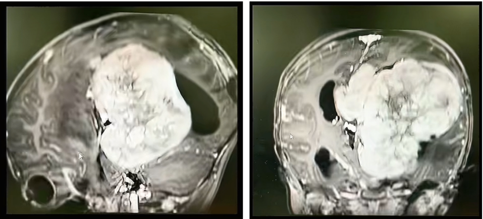
Fig. 2 Preoperative imaging of the patient, showing the giant intracranial tumor.
The operation was only the beginning
Postoperative recovery initially went well. The child woke up, regained stable breathing, preserved full limb strength, and showed no evident language deficit. But about a week later, she developed a very severe intracranial infection, with cerebrospinal fluid white blood cell counts rising dramatically and then rebounding even after antibiotic treatment had begun.
Gao said this was exactly why neurosurgeons cannot focus on the operation alone. After giant intracranial tumors are removed, a cavity remains that can collect fluid and blood, creating a fertile environment for infection inside a closed compartment where treatment is difficult. He has spent decades building an integrated neurocritical care model that extends from the operating room into postoperative rescue and long-term management.
In this case, the team used culture-guided anti-infective therapy, intrathecal antibiotic delivery, daily controlled cerebrospinal fluid drainage through a lumbar catheter, and repeated expert consultations to adjust treatment. Those efforts gradually brought the infection under control and restored the cerebrospinal fluid parameters to normal.
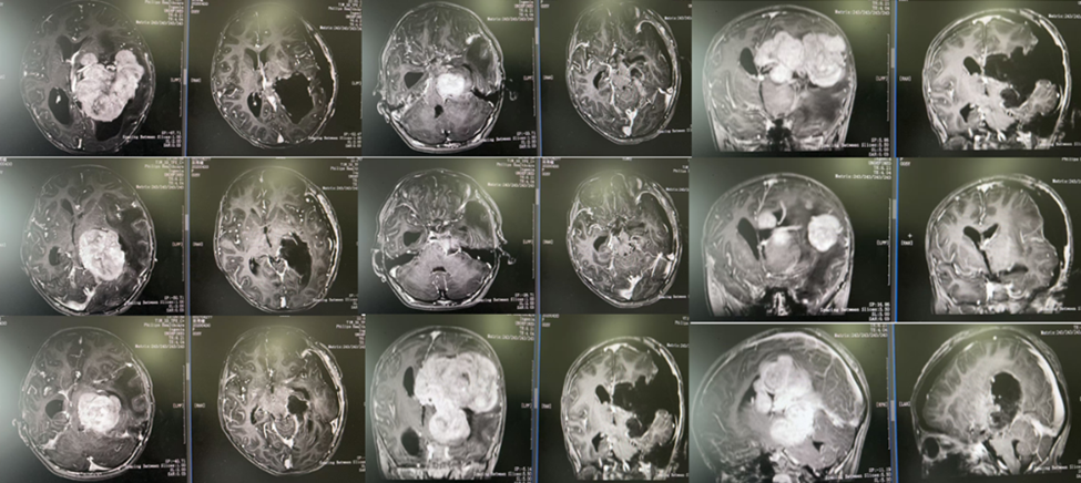
Fig. 3 Pre- and postoperative imaging comparison from the original article.
A benign diagnosis and a new life ahead
Pathology ultimately confirmed a World Health Organization grade 1 meningioma, a benign tumor with a very low risk of recurrence. By discharge, the previously displaced midbrain had returned toward a normal position, two thirds of the intracranial cavity had already refilled, and the team expected that the residual space would continue to resolve over the next few years.
According to Gao, the child was discharged with intact limb movement, fluent language, and quick thinking, with no epilepsy or other major complications. She will continue with follow-up and electroencephalographic monitoring, but her doctors expect her to study, live, and grow like other healthy children.
For Gao, the success of the case came from three elements working together: refined neurosurgical technique, tightly organized neurocritical care, and seamless cross-institutional collaboration between a specialty neurological center and a pediatric hospital. The result was not just a difficult tumor resection, but the restoration of a life that many had already given up on.
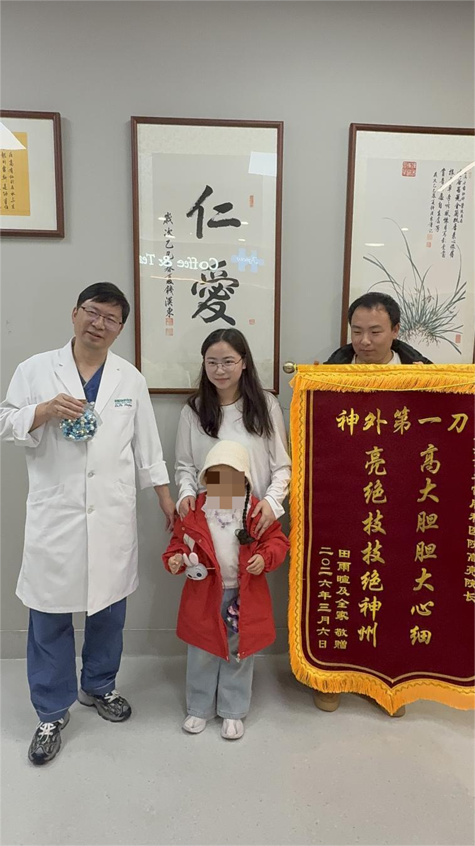
Fig. 4 The patient and her family with the medical team at discharge.
.

