


Background and Unmet Need
Diffuse large B‑cell lymphoma (DLBCL) exhibits prominent heterogeneity in tumour cells and the immune microenvironment. Conventional bulk‑RNA sequencing only yields aggregate gene‑expression profiles. While single‑cell sequencing permits characterisation of cell types and cellular states, it cannot preserve the spatial locations of cells. Reliance merely on immune‑cell abundance fails to account for disparities in treatment response and patient survival.
Recently, a spatial transcriptomics study published in OncoImmunology reconstructed the DLBCL tumour‑microenvironment framework from multiple dimensions including spatial organisation, cellular interactions and transcriptional functions, and validated its prognostic value in an independent cohort comprising more than 1,000 cases. Professor Zhou Hui and Dr. Wang Caiqin from Hunan Cancer Hospital have been invited in this article to interpret the research.
Expert profile
Zhou Hui

· Doctor of Oncology, Professor, Chief Physician, Master's Supervisor
· Member of the Party Committee and Vice President of Hunan Cancer Hospital
· Member of the Lymphoma Quality Control Expert Committee of the National Cancer Quality Control Center
· Deputy Chair Member, Lymphoma Committee of China Anti Cancer Association
· Deputy Chair Member, Lymphoma Integrated Rehabilitation Committee of China Ant Cancer Association
· Director of Hunan Provincial Lymphoma Diagnosis and Treatment Center
· Director of Hunan Provincial Engineering Research Center for Precision Diagnosis and Treatment of Lymphoma
· Member, Lymphoma Group, Oncology Branch of Chinese Medical Association
· Member, Cancer Translational Medicine Group, Oncology Branch of Chinese Medical Association
· Standing Committee Member, Lymphoma Expert Committee of CSCO
· Candidate Chair Member, Medical Oncology Committee of Hunan Medical Association
Wang Caiqin

· Attending Physician, Department of Lymphoma Hematology, Hunan Cancer Hospital
· Main research interest: mechanisms and clinical translational research of drugs enhancing antitumor immune responses
· First/corresponding author of multiple papers published on Journal for ImmunoTherapy of Cancer, Cancer Communications, Journal of Advanced Research, Journal of Translational Medicine, among which 3 papers have an impact factor over 10
· 1 authorized invention patent as the first inventor
· Principal investigator for 1 national level, 1 provincial level and 1 post doctoral startup fund projects
· Committee Member, Tumor Immunotherapy Committee of China Elderly Health Care Association
· Committee Member, Stemcell and CART Cell Therapy Branch, Guangdong Health Popularization Promotion Association
Study Design
The discovery cohort combined spatial transcriptomics with reference-based deconvolution:
· 10x Genomics Visium spatial transcriptomics (55-micrometer spots) on 10 formalin-fixed paraffin-embedded (FFPE) tissue sections from 8 DLBCL patients.
· Section breakdown: 6 pre-treatment sections from different patients; 2 longitudinal pre- and post-treatment sections from 1 patient; 2 pre-treatment sections from 2 distinct tumor sites in 1 patient.
· 4 reactive lymph node and tonsil sections as secondary lymphoid-organ controls.
· Reference-based deconvolution with the RCTD method against published DLBCL single-cell transcriptomes to estimate cell-type composition of each spot.
· Unsupervised Leiden clustering of spot-level compositions to identify recurring local communities, labeled as spatial cell ecosystems (Cell-Ecos).
· Neighborhood scoring, differential expression analysis, pathway enrichment, Moran's I spatial autocorrelation, and ICELLNET ligand-receptor communication analyses.
· Multiplex immunofluorescence for CD20, CD3, and CD68 as orthogonal spatial validation.
· Survival validation in 1,149 DLBCL patients from the Hematological Malignancy Research Network (HMRN), with the Visco cohort used for cross-validation.
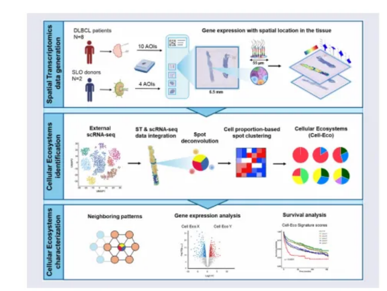 Figure.1.Complete Technical Flowchart of the Study
What the Study Found
Eleven core cell types were resolved, including malignant B cells, non-malignant B cells, macrophages, CD4+ and CD8+ T cells, regulatory T cells (Treg), T follicular helper cells (Tfh), natural killer (NK) cells, conventional dendritic cells (cDCs), plasmacytoid dendritic cells (pDCs), and plasma cells. The proportion of malignant B cells correlated inversely with overall immune infiltration. In low- and moderate-infiltration samples, CD8+ T cells and macrophages co-localized and correlated negatively with malignant B cells; high-infiltration samples were enriched for CD4+ T cells with different spatial co-occurrence patterns.
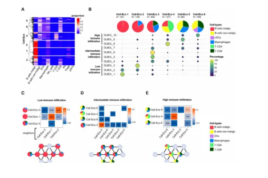 Figure.2.Composition, Sample Distribution and Neighborhood Structure of Six Categories of Cell Eco
Six stable Cell-Ecos emerged across samples: (1) malignant-B-cell dominant; (2) malignant B cells with macrophages; (3) malignant B cells with macrophages and CD8+ T cells; (4) malignant B cells with non-malignant B cells and CD4+ T cells, approximating a normal lymph-node-like structure; (5) non-malignant B cells with macrophages and CD8+ T cells; and (6) non-malignant B cells with macrophages, CD8+ T cells, and cDCs, the most immunologically diverse ecosystem. Low-infiltration samples were enriched for Cell-Ecos 1 and 2 forming tumor-cell-rich spatial aggregates, whereas moderate- and high-infiltration samples showed more dispersed Cell-Ecos 5 and 6.
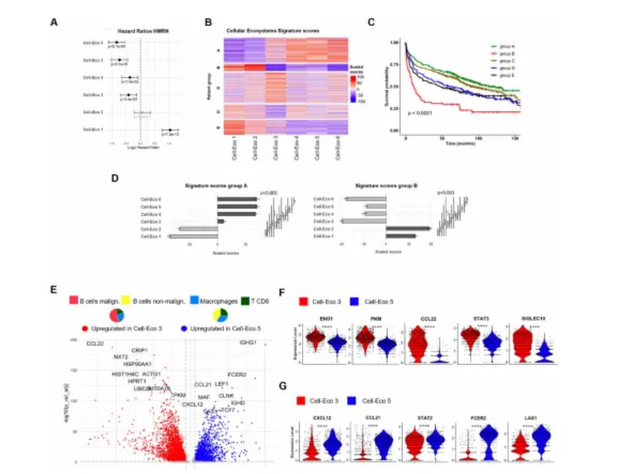 Figure.3.Prognostic Stratification of Cell Eco and Functional Differences Between Cell Eco 3 and Cell Eco 5
Why It Matters
A Cell-Eco-derived gene signature applied to the 1,149-patient HMRN cohort separated patients into five groups with significantly different overall survival (log-rank P<0.0001). Patients with high immune-ecosystem scores (Cell-Ecos 4-6) had the most favorable long-term outcomes, while those with high malignant-ecosystem scores (Cell-Ecos 1-2) and low immune-ecosystem scores had the worst. The signature aligned with, but was not fully explained by, COO, LymphGen subtype, or IPI, suggesting that spatial ecosystem features may add a microenvironmental prognostic dimension to established classifiers.
Mechanistic Insights
Two paired comparisons show why composition alone can mislead.
Cell-Eco 3 vs Cell-Eco 5. Both contain CD8+ T cells and macrophages, yet Cell-Eco 3 also contains malignant B cells, shows strong glycolysis and proliferation programs, and up-regulates ENO1, PKM, CCL22, STAT3, and SIGLEC10/CD24, consistent with an immune-suppressed, metabolically active niche. Cell-Eco 5 is dominated by non-malignant B cells and instead expresses CXCL12, CCL19, CCL21, and STAT2/STAT4, suggesting active immune-cell recruitment and T-cell functional programs.
Cell-Eco 1 vs Cell-Eco 2. Both are malignant-B-cell rich, but Cell-Eco 2 is enriched for macrophage genes including C1QC, APOE, CD74, and IL4I1, consistent with C1q-positive macrophage features involved in antigen presentation and metabolic reprogramming. Ligand-receptor analyses highlighted LGALS1-LGALS1, APP-CD74, and CXCL12-CCR4 pairs, which may dampen macrophage anti-tumor activity or foster tumor-supportive crosstalk. In low-infiltration samples, 39 spatially auto-correlated genes tracked macrophage proportions rather than malignant B-cell proportions, supporting macrophages as local organizers of tissue remodeling. In one longitudinal case, the proportion of Cell-Eco 2 rose from 41.08% to 86.24% at relapse, hinting at ecosystem remodeling after R-CHOP in a single patient.
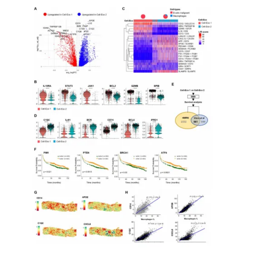 Figure.4.Macrophage Associated Programs, Cell-Cell Communication and Spatial Organizational Features within the Malignant B-cell Ecosystem
Translational Value
The findings provide a rationale for evaluating spatial ecosystem features as candidate prognostic biomarkers beyond COO, IPI, and LymphGen, and for targeting macrophage-organized niches. Candidate intervention axes emerging from the analysis include the C1q/complement pathway, CCL22, CD24-SIGLEC10, STAT3 signaling, glycolysis, and selected ligand-receptor pairs such as LGALS1-LGALS1, APP-CD74, and CXCL12-CCR4. The work supports further evaluation in larger, uniformly treated cohorts and integration with single-cell spatial platforms, digital pathology, and circulating tumor DNA (ctDNA) monitoring.
Clinical Evidence Status
This is a translational research study using spatial transcriptomics in a small discovery cohort and a single retrospective observational validation cohort. No prospective clinical trial, no patient-level clinical intervention, and no outcome data derived from acting on Cell-Eco classification are reported. Any clinical application of these findings remains hypothetical and requires prospective validation.
Limitations and Next Steps
Several limitations should temper interpretation:
· Discovery was based on 10 sections from 8 patients, with intra-patient replicates that may inflate apparent independence.
· Each 55-micrometer spot contains multiple cells, so direct cell-cell contact relationships were inferred rather than measured.
· Bulk RNA-seq in the validation cohort cannot confirm that the same spatial structures exist in those patients.
· Fibroblasts and additional stromal components were excluded because of single-cell reference limits.
· Post-treatment ecosystem remodeling is supported by a single longitudinal case.
· Larger, fully annotated, uniformly treated cohorts with single-cell resolution spatial profiling, digital pathology, and ctDNA monitoring are needed to assess reproducibility and treatment-predictive value.

