


The third plenary session of the 46th World Congress of the Société Internationale d’Urologie (SIU) brought together six reports on clinical practice and technology in urology. The session, chaired in honour by Prof. Jean de la Rosette of Istanbul Medipol University and Prof. Yasser Farahat of Tanta University, Egypt, covered affordable urological innovation, prostate cancer risk factors and stratified management, value-based urology, robot-assisted ureteric reconstruction and new techniques in stone treatment.
Article Key Points
• Six reports covered affordable training, prostate cancer risk, value-based care and stone treatment
• PCNL puncture training models were built from mobile phones, gloves and ordinary packaging boxes
• PSMA-PET risk stratification may spare low-risk patients unnecessary extended lymph node dissection
• The planned APPLY-PSMA (S2613) study compares omission, targeted and extended dissection by PSMA-PET result
• Endoscopic practice is moving from fragmentation towards active clearance with suction and pressure monitoring
Expanding Training and Clinical Practice from Existing Resources
In regions where healthcare resources are limited, running surgical training and solving real clinical problems with the conditions at hand is a practical challenge that urologists face. Prof. Mohammed Lezrek described his experience of running urological clinical training in several African countries, and shared practices that developed out of resource-limited settings.
Lezrek pointed out that innovation does not necessarily depend on new equipment, and can instead begin with existing instruments and training methods. In his report he described building PCNL puncture training models from mobile phones, gloves and ordinary packaging boxes, practising tract dilation with watermelons and pumpkins, and running simulation training for TURP and partial nephrectomy using animal kidneys. He also shared the experience of meeting some intraoperative instrument needs with common materials such as hydrophilic guidewires, nylon suture and metal cores for ureteric catheters.
These practices illustrate a different approach to affordable innovation. Starting from real clinical problems and making flexible use of what is already available, it is still possible to run surgical training and improve technique, and to offer more options for urological practice in resource-limited settings.
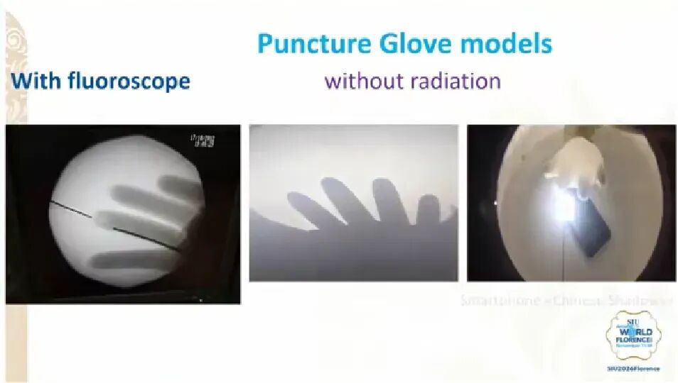 Fig. 1. Prof. Mohammed Lezrek Demonstrating the Puncture Training Model
Testosterone, Genetics and Environmental Factors: A Multidimensional Exploration of Prostate Cancer Risk
Prostate cancer develops in relation to a range of factors, including hormone levels, genetic background and environmental influences, and the picture may differ between populations. Prof. Shingai Mutambirwa approached the question through research in African populations, and described findings on the relationship between testosterone and prostate cancer as well as the questions that warrant further study.
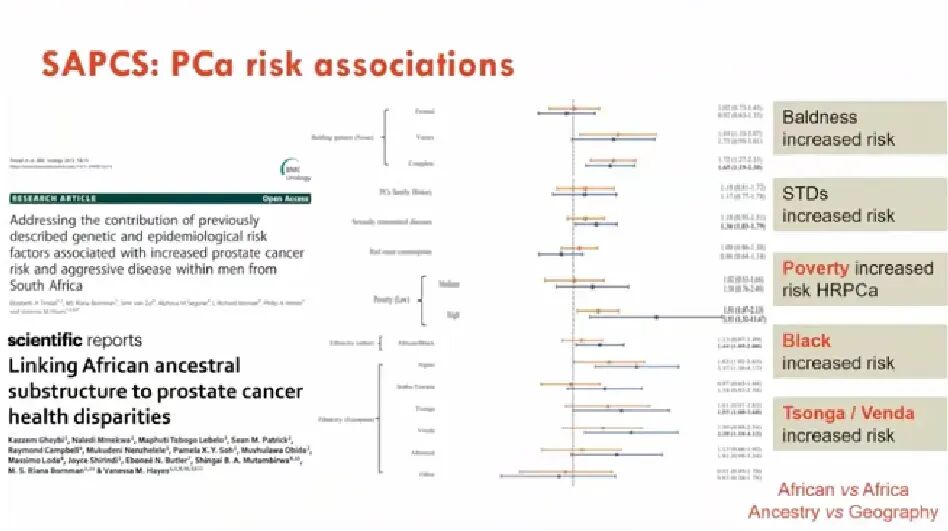 Fig. 2. Prof. Shingai Mutambirwa Presenting on the Associations of Prostate Cancer Risk
In his report, Mutambirwa drew particular attention to the distinction between total and free testosterone, and set out the view that free testosterone may be more closely related to prostate cancer risk. Differences in incidence between different population groups in South Africa also point to a possible role for genetic background. Beyond hormonal and genetic factors, environmental and biological factors such as DDT exposure and changes in the microbiome are directions that warrant further exploration.
These studies also expose a problem in current prostate cancer research: much of the available evidence comes from European and North American populations, and African population groups remain insufficiently characterised. Mutambirwa’s view is that research within African populations should be strengthened, accumulating data that are regionally and ethnically representative, so that prostate cancer risk factors and their population differences can be described more accurately.
Balancing Efficacy and Cost: Value-Based Urology
As robotic surgery, laser therapy and other new technologies are applied more widely in urology, how to improve treatment outcomes while also accounting for healthcare costs has become a question for clinical practice. Prof. Samuel Vincent G. Yrastorza drew on clinical experience from different institutions to share his understanding of value-based urology. His view was that technology choice cannot rest on whether a technique is advanced, but must also take account of patient need, economic burden and the actual conditions of care.
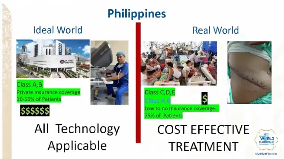 Fig. 3. Prof. Samuel Vincent G. Yrastorza Sharing Practical Experience of Value-based Urology
With robotic surgery as an example, robot-assisted radical prostatectomy and complex partial nephrectomy have certain advantages, but for relatively straightforward operations the decision to use a robot still has to be made on the individual case. Treatment of benign prostatic hyperplasia should likewise be chosen between laser enucleation and conventional TURP according to prostate volume and the patient’s condition.
Yrastorza also pointed out that the development of new techniques should not come at the cost of neglecting conventional operations. Urologists still need to master several surgical approaches, so that treatment can be matched to the patient and the available resources. Technology is not the end in itself; ensuring that patients derive real benefit is the core concern of value-based care.
From Routine Dissection to Risk Stratification: Exploring Pelvic Lymph Node Dissection Strategies in Prostate Cancer
For patients undergoing radical prostatectomy, whether pelvic lymph node dissection is necessary, and what extent of dissection should be used, has long been a focus of clinical debate. In this session, Prof. Udit Singhal drew on existing research evidence to consider how PSMA-PET can be used to stratify patients by risk, avoiding unnecessary dissection in low-risk patients while ensuring that those who genuinely need it are adequately treated.
Singhal pointed out that the risk of nodal metastasis differs between patients with unfavourable intermediate-risk and high-risk prostate cancer, so a uniform surgical strategy cannot be based on clinical risk groups alone. Randomised studies, including those from the teams of Lestingi and Touijer, have not yet demonstrated a clear benefit of extended pelvic lymph node dissection on primary endpoints, while the dissection itself may bring complications such as lymphoedema, lymphocele and thromboembolism. With PSMA-PET increasingly applied in clinical practice, combining imaging results with clinical risk prediction models may allow patients with a low risk of nodal metastasis to be identified more accurately, providing a reference for individualising the extent of dissection.
Singhal also introduced the APPLY-PSMA (S2613) study, which is in preparation. The study stratifies patients according to PSMA-PET results: for imaging-negative patients it compares omission of lymph node dissection with extended dissection, and for patients with 1-2 positive lymph nodes it compares targeted dissection with extended dissection, with metastasis-free survival as the primary endpoint, to assess the feasibility of adjusting the extent of dissection under PSMA-PET guidance. The study will also explore how surgical strategy can be set according to a patient’s actual risk of metastasis, avoiding unnecessary dissection while also accounting for tumour control.
 Fig. 4. Study Design of APPLY-PSMA (S2613)
Surgical Choice Depends on the Lesion: Selecting the Technique in Robotic Ureteric Reconstruction
Ureteric reconstruction involves several types of lesion, and because the site of the stenosis, the length of the defect and the condition of the surrounding tissue differ, the surgical approach varies accordingly. Prof. Devang Desai used clinical cases of robot-assisted reconstruction to describe the management of pelviureteric junction obstruction, ureteric stricture and complex recurrent disease, and shared his experience of protecting ureteric tissue and its blood supply during reconstruction.
For complex or recurrent strictures, Desai introduced buccal mucosa graft patch augmentation and appendiceal patch reconstruction. Compared with excising the diseased segment and substituting bowel, when conditions allow, augmenting the existing ureter with a patch reduces tissue excision and additional injury. The view afforded by robotic surgery and the availability of fluorescence imaging also assist in identifying anatomy, observing tissue perfusion and assessing the anastomosis during the operation. For distal ureteric lesions, the choice between bladder suspension and Boari flap plasty depends on bladder function and the length of the defect.
Desai also described progress in research on tissue engineering and decellularised matrix biomaterials for reconstruction of the urinary tract. Lesions at different sites and of different types require corresponding reconstructive approaches, and the operating plan should be set according to the individual case. Preserving tissue viability and local blood supply remains an important determinant of the outcome of reconstruction.
Beyond Fragmentation: Endoscopic Urology Enters the Era of Active Stone Clearance
With the development of flexible ureteroscopes, laser technology and suction systems, the goal of endoscopic urologic surgery is shifting from simply fragmenting stones to actively removing them. Prof. Bogdan Geavlete pointed out that genuinely transformative technology has to improve operative efficiency while also accounting for safety and patient benefit.
The report focused on four areas of technical progress. First, laser technology is moving from pure energy delivery towards intelligent functions such as tissue and stone recognition. Second, miniaturisation of ureteroscopes may reduce ureteric injury and improve access to the calyces, although instrument diameter still has to be balanced against irrigation flow and working channel performance. Third, suction technology, through approaches such as the flexible and navigable suction ureteral access sheath (FANS) and direct in-scope suction, removes fragments during fragmentation, improving the visual field and helping to lower intrarenal pressure. Fourth, intelligent pressure control monitors intrarenal pressure in real time and coordinates irrigation with suction, further improving procedural safety.
Geavlete’s view was that the key to the future of endoscopic urology is not simply a thinner ureteroscope, a more powerful laser or a more efficient suction device, but the integration of instruments, energy, active clearance and real-time monitoring, moving surgery towards an intelligent and controllable model of active stone clearance.
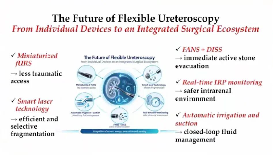 Fig. 5. Prof. Bogdan Geavlete on the Future Trend of Integrated Endourological Devices
Summary
This session spanned clinical practice and technical advances across several areas of urology, from clinical innovation in resource-limited settings to prostate cancer risk assessment and surgical decision-making, and on to robot-assisted ureteric reconstruction and endoscopic stone treatment technology. The contributions were a mix of re-examination of current approaches and exploration of new technologies in clinical application. Whether the surgical method is chosen according to the individual patient, or treatment decisions are refined with imaging and new devices, both ultimately return to clinical need and patient benefit. As the relevant research and technologies continue to develop, how best to apply these advances to daily practice remains a question for urologists to follow.

