


This article was authored and reviewed by Prof. Ling Li, Chief Physician and Master’s Supervisor at Jiangxi Maternal and Child Health Hospital, whose research focus centers on gynecologic radiation oncology and brachytherapy.
Expert Profile: Prof. Ling Li
- l Chief Physician, Master’s Supervisor; Director, Radiotherapy Center for Oncology, Jiangxi Maternal and Child Health Hospital
- l Vice Chair, Committee on Gynecologic Oncology Prevention and Treatment, Chinese Maternal and Child Health Association
- l Standing Member, Youth Council, China Anti-Cancer Association (CACA)
- l Standing Committee Member, Brachytherapy Committee, China Anti-Cancer Association
- l Chair, Brachytherapy Committee, Jiangxi Anti-Cancer Association
- l Chair-elect, Cervical Cancer Prevention and Treatment Committee, Jiangxi Maternal and Child Health Association
- l Selected for the Jiangxi Province “Hundreds, Thousands, Tens of Thousands” Talent Program
- l Visiting Scholar, U.S. National Cancer Institute (NCI)
- l Recipient of the Second Prize, Chinese Maternal and Child Health Science and Technology Achievement Award; Third Prize, Science and Technology Award, China Anti-Cancer Association; Second Prize, Jiangxi Province Science and Technology Progress Award
- l Named “National Famous Doctor: Young and Emerging Talent” by People’s Daily Online in 2019
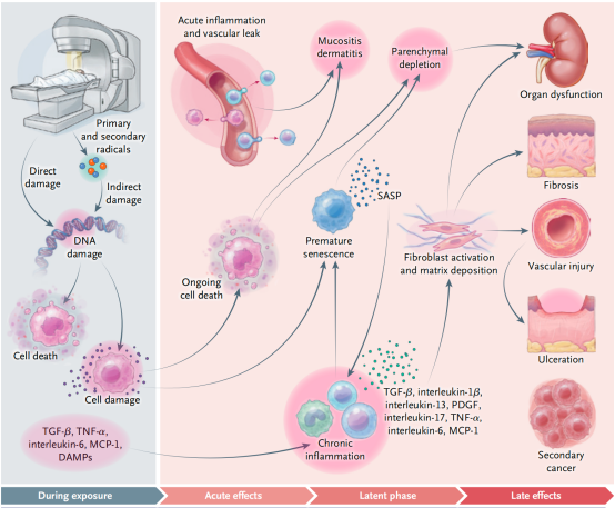
A recent review in The New England Journal of Medicine titled “Effects of Radiotherapy in Normal Tissue” systematically catalogues what happens when ionizing radiation enters normal tissue: DNA damage, free-radical formation, inflammatory response, vascular changes, stem-cell aging, parenchymal cell loss, and long-term fibrosis.
But reading this review, I was drawn back to an old question in cervical cancer radiotherapy: hypoxia.
Because radiotherapy is never simply a question of “how much dose.” It is always a balance between two biological worlds: the tumor that must be eliminated and the normal tissue that must be preserved as much as possible. And oxygen plays a complex, sometimes contradictory, role in both.
Why Hypoxia Can Influence Radiotherapy
One important mechanism by which radiation kills cells is by inflicting DNA damage. Oxygen can participate in “fixing” radiation-induced DNA damage, making the lesion harder for the cell to repair.
In well-oxygenated tumor cells, radiation injury is therefore generally more thorough; in hypoxic environments, some of the damage can be repaired and tumor cells exhibit stronger radioresistance. This is the classical “oxygen effect.”
But the consequences of hypoxia go well beyond this. Under low-oxygen pressure, tumor cells activate HIF-related pathways, increase VEGF expression, alter glucose metabolism, switch on autophagy and stress responses, and progressively acquire stronger invasive, metastatic, and survival capacities.
So hypoxia does not merely let radiation “kill a few cells fewer.” It may instead select for a subpopulation of tumor cells that is more treatment-tolerant and more invasive.
Cervical cancer is among the most classic tumor types for hypoxia research. In the clinic, large-volume tumors with obvious necrosis, nodal metastases, or associated hydronephrosis routinely alert us to more severe perfusion insufficiency and hypoxic microenvironment behind them.
A 55-Gene Study That Identifies True “Hypoxic Cervical Cancer”
In 2026, a cervical cancer hypoxia study was published in Radiotherapy and Oncology. The core question the investigators wanted to answer was: can we use pre-treatment tumor biopsies to identify truly “hypoxic cervical cancer”?
The investigators first cultured five cervical cancer cell lines under 21% oxygen (normoxia) and 1% oxygen (hypoxia) for 24 hours, then used RNA sequencing to identify genes that consistently respond to hypoxia.
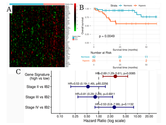
The study identified 402 differentially expressed genes, narrowed these to 61 candidates that changed in the same direction in at least four of the five cell lines, and further matched 55 of these against TCGA cervical cancer data—producing the “Manchester 55-gene hypoxia signature.”
All 55 genes were upregulated under hypoxia. They include CA9, VEGFA, SLC2A1, HK2, LDHA, PGK1, BNIP3, and EGLN1, among others, and span hypoxia response, angiogenesis, glycolysis, metabolic reprogramming, and cellular stress, providing a coherent biological rationale.
The investigators then validated this gene signature in multiple patient cohorts.
In the TCGA cohort, hypoxic patients had worse overall survival and progression-free survival. After adjustment for clinical stage, hypoxia classification remained an independent adverse factor for overall survival, with HR 2.69.
In the Manchester cohort of 153 patients, hypoxic tumors were more common in patients with later-stage disease, tumors at least 4 cm in diameter, pelvic nodal metastases, and hydronephrosis. In multivariable analysis, the hypoxia signature remained independently associated with worse overall survival, HR 1.95.
These findings were then replicated in Korean and Norwegian cohorts.
In other words, the 55 genes identify not merely a laboratory phenomenon of hypoxic response, but a clinical phenotype tied to tumor burden, radioresistance, and patient prognosis.
“Poor Effect” Does Not Mean “Escalate the Dose”
The 55-gene signature is currently a prognostic biomarker, not a predictive one. What it tells us is that these patients are at higher risk.
It does not tell us: should the external-beam dose be increased? Should the fractionation schedule be altered? Should the brachytherapy dose be increased? Should a hypoxia-targeting radiosensitizer be added? Are these patients better served by immunotherapy?
Without prospective interventional trials, blindly escalating dose simply because a tumor is “hypoxic” is unlikely to solve the problem and may instead increase injury to the bladder, rectum, sigmoid colon, small bowel, and bone marrow.
This is precisely the reminder embedded in the NEJM review: when we think about overcoming tumor resistance, we must not forget that normal tissue has its own radiobiology.
Normal-Tissue Tolerance Limits Are Not a Simple Table
The NEJM review notes that radiation-induced normal-tissue injury is not a single event that appears suddenly at the end of treatment, but a dynamic process that may continue for months or even years.
In cervical cancer treatment, the acute diarrhea, bladder irritation, and bone-marrow suppression that we monitor, and the late rectal bleeding, bladder injury, vaginal stenosis, pelvic fibrosis, and lymphedema that we strive to prevent, cannot be explained by a single “exceeded the tolerance dose” threshold.
Radiation dose, dose per fraction, irradiated volume, concurrent chemotherapy, prior surgery, underlying comorbidities, and individual radiosensitivity together determine the normal-tissue outcome.
A Personal View
Hypoxia-gene research is genuinely attractive, but the most reliable route to better outcomes in cervical cancer today is still to deliver standard treatment completely and to specification:
- Complete external-beam radiotherapy and platinum-based concurrent chemotherapy as prescribed;
- Individualize dose escalation to involved nodes;
- Complete image-guided adaptive brachytherapy;
- Use intracavitary combined with interstitial implantation when indicated;
- Ensure the high-risk clinical target volume receives adequate cumulative EQD2;
- And concurrently keep bladder, rectum, sigmoid, and small-bowel doses within tolerance while avoiding unnecessary treatment interruptions and total treatment-time prolongation.
Because hypoxic tumors are already radioresistant. If treatment time is further extended due to acute toxicity, planning gaps, or limited brachytherapy resources, this may give the residual tumor cells additional time to repopulate.
In other words, until we can precisely reverse hypoxia, we cannot afford to let treatment quality compound the problem that hypoxia already creates.
The future of radiotherapy is not only about aiming more precisely at the tumor; it is about more accurately understanding the biology of both tumor and normal tissue.
Moving from “one‑dose‑fits‑all” to biologically individualized radiotherapy is, perhaps, exactly what precision radiotherapy ought to achieve.

