


A 6-centimeter fetus implanted in the liver—a medical rarity—was recently diagnosed and successfully treated at Xi’an People’s Hospital (Xi’an Fourth Hospital). The case marks the hospital’s first documented hepatic pregnancy, according to Dr. Biliang Chen, Director of the Obstetrics and Gynecology Hospital.
Ectopic pregnancy is a common gynecologic emergency, with more than 95% occurring in the fallopian tubes. In contrast, hepatic pregnancy—implantation of a fertilized egg on or within the liver—is exceptionally rare. Fewer than 50 cases have been reported worldwide, with an estimated incidence of approximately 1 in 15,000 intrauterine pregnancies.
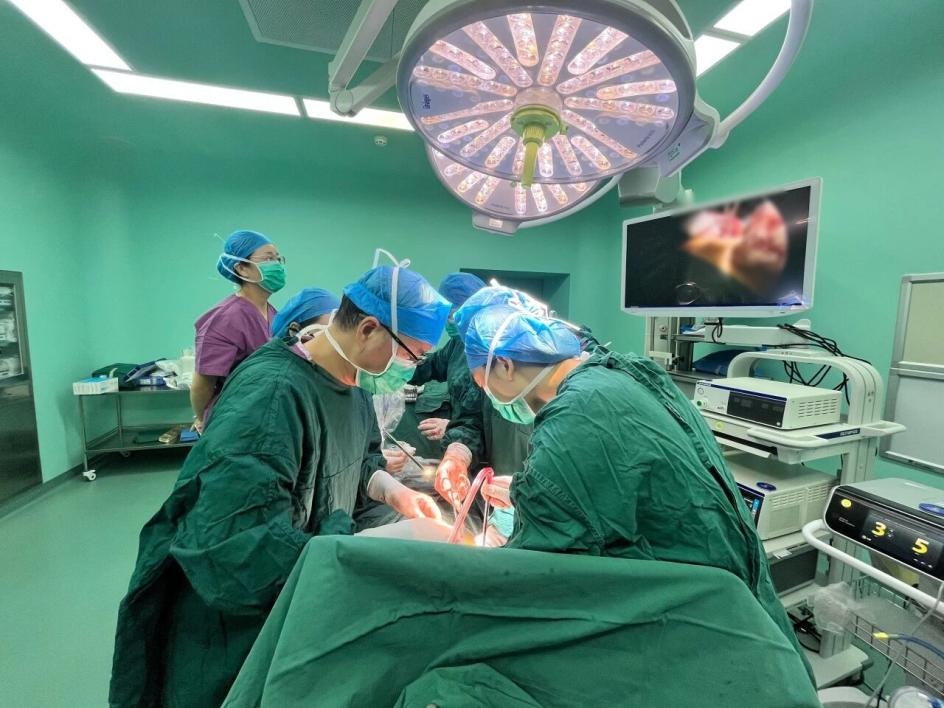
The surgical procedure was performed for the patient by the team led by President Biliang Chen in collaboration with experts from the Department of Hepatobiliary Surgery. / All images in this article are sourced from Xi'an People's Hospital.
The patient, a 40-year-old woman identified as Ms. Wang (pseudonym), presented with amenorrhea, nausea, and vomiting. Blood tests confirmed pregnancy, with an abnormally high HCG level of approximately 90,000 mIU/mL, yet repeated ultrasound examinations failed to locate a gestational sac in the uterus or fallopian tubes. She had a prior history of tubal pregnancy and left salpingectomy.
After transfer to Xi’an People’s Hospital, further evaluation was conducted. A meticulous ultrasound examination by Dr. Sanli Guan from the Department of Ultrasound ultimately identified a fetus within the liver. The crown–rump length measured approximately 3.6 cm, corresponding to a gestational age of nearly 11 weeks, with detectable fetal cardiac activity. The preliminary diagnosis was hepatic pregnancy.
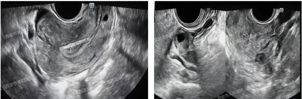
The left image shows no gestational sac visible in the uterine cavity on intracavitary ultrasound. The right image reveals a small cyst in the right ovary.
Subsequent magnetic resonance imaging (MRI) localized the pregnancy to segment VI (S6) of the liver. Based on clinical findings, imaging, and exclusion of secondary implantation, the patient was diagnosed with primary hepatic pregnancy—the rarest form of abdominal pregnancy.
Given the advanced gestational age and the liver’s rich vascular supply, conservative treatment with methotrexate was deemed unsafe. A multidisciplinary team involving obstetrics, hepatobiliary surgery, anesthesiology, and transfusion services determined that partial hepatectomy offered the safest therapeutic option.
The surgical team performed an open partial hepatectomy of liver segment S6 via a right upper quadrant L-shaped incision. The operation lasted over two hours and was completed successfully. Intraoperative measurements showed the fetus measured nearly 6 cm. The patient recovered uneventfully and was discharged with near-normal postoperative HCG levels.
Dr. Chen emphasized that the primary diagnostic challenge of hepatic pregnancy lies in its rarity. Elevated HCG levels without identifiable intrauterine or tubal gestation should prompt clinicians to expand the diagnostic field beyond the pelvis.
Early detection is critical. Rupture of a hepatic pregnancy carries a mortality risk more than seven times higher than that of ruptured tubal pregnancy. Dr. Chen noted that most reported cases worldwide were diagnosed only after catastrophic bleeding occurred.
Recent studies suggest that the incidence of ectopic pregnancy in China is increasing, including implantation at rare sites such as the liver and spleen. Risk factors may include tubal disease, pelvic inflammation, contraceptive methods, and assisted reproductive technologies.
Dr. Chen advises women of reproductive age—particularly those with a history of ectopic pregnancy—to seek immediate medical evaluation for symptoms such as amenorrhea, abdominal pain, or vaginal bleeding, stressing that early diagnosis can be lifesaving.

