


Spina bifida, a congenital defect and one of the most severe neural tube defects, places a heavy health and economic burden on families and society. Although preventive measures such as periconceptional folic acid supplementation are widely implemented, many affected children still face lifelong complications including paralysis, bowel and bladder dysfunction, and hydrocephalus, requiring long-term multidisciplinary care. Previously, fetal surgical repair could reduce the incidence of hydrocephalus and improve some motor function, but most children still could not walk independently. Recently, the first-in-human clinical trial (CuRe trial) published in The Lancet achieved a breakthrough, demonstrating that the combination of placental mesenchymal stromal cells (PMSCs) with fetal surgery for myelomeningocele repair is safe and feasible, opening a new treatment path for this congenital disease.
Fig. 1. Screenshot of the published article.
The Challenge and Exploration: Limitations of Traditional Treatment and the Rise of Stem Cell Therapy
Myelomeningocele is the most severe form of spina bifida, resulting from incomplete neural tube closure in early embryonic development, which leads to the protrusion of the spinal cord and meninges through a vertebral defect, exposing them to amniotic fluid. This exposure ultimately leads to a series of severe consequences, including lower limb paralysis, bladder and bowel dysfunction, and Arnold-Chiari type II malformation with hindbrain herniation. Traditional surgery can only achieve mechanical closure and cannot reverse the spinal cord injury that has already occurred. Therefore, innovative therapeutic approaches with both repair and regenerative functions are urgently needed.
The neuro-reparative potential of mesenchymal stromal cells offers a direction to address this challenge. PMSCs stand out due to their unique advantages: as fetal-derived cells, they are non-tumorigenic and can secrete various neurotrophic, anti-inflammatory, and anti-apoptotic factors. They also possess immunomodulatory capabilities, promoting neurite outgrowth and protecting damaged neurons. Previous studies in a sheep model confirmed that fetal repair using PMSCs seeded on an extracellular matrix significantly improved motor function and restored normal bowel and bladder function, laying a solid foundation for conducting human clinical trials.
In-Depth Analysis of the CuRe Trial: Design and Key Findings of the First-in-Human Study
The CuRe trial is a phase 1, first-in-human, single-center, single-arm study designed to evaluate the safety and feasibility of a placental mesenchymal stromal cell-extracellular matrix (PMSC-ECM) construct for fetal myelomeningocele repair. The study enrolled six pregnant women between June 2021 and December 2022. Fetal gestational age ranged from 24 weeks + 5 days to 25 weeks + 5 days. The upper boundary of the myelomeningocele defect was located between the T1 and S1 vertebral segments. MRI showed hindbrain herniation, and the karyotype was normal. The primary safety outcomes included assessing the healing of the repair site, and the occurrence of cerebrospinal fluid leaks, infections, abnormal growths, or tumor formation.
Core Study Results
Surgery was successfully completed in all six fetuses without intraoperative complications. The PMSC-ECM construct was successfully applied to the defect site in all cases, confirming the technical feasibility of the therapy. All newborns were delivered by cesarean section at a median gestational age of 34 weeks + 5 days (range: 33 weeks + 2 days to 36 weeks + 6 days). One infant required intubation for respiratory distress syndrome due to prematurity, but all infants were discharged without requiring home oxygen therapy.
Key safety indicators met expectations: All neonates had intact repair sites at birth, with no cerebrospinal fluid leaks, infections, or abnormal tissue growth. MRI examinations within the first two weeks after birth showed complete reversal of hindbrain herniation in all infants and no abnormal tissue proliferation or tumor formation. None of the infants required a ventriculoperitoneal shunt for hydrocephalus before discharge. Follow-up data as of August 1, 2025, showed no stem cell-related wound complications, further validating the safety of the therapy.
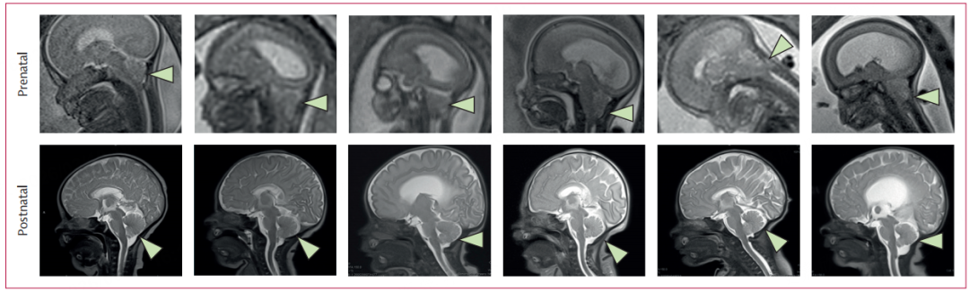
Fig. 2. Prenatal and postnatal MRI imaging results.
Compared to historical data from the MOMS trial, the baseline characteristics of the fetuses in this study (such as gestational age at surgery and lesion level distribution) were comparable, and no additional safety risks were observed, indicating that the PMSC-ECM construct is compatible with standard fetal surgery and does not interfere with the benefits of the original treatment.
Clinical Significance and Future Perspectives
As the first human study to evaluate fetal stem cell therapy for myelomeningocele, the CuRe trial has significance far beyond mere safety verification. This study is the first to demonstrate that allogeneic PMSCs can be safely applied to the fetal central nervous system, addressing long-standing concerns about fetal tolerance to allogeneic stem cells and laying an important foundation for the development of fetal regenerative medicine.
The research team has already initiated a phase I/IIa clinical trial, planning to enroll 35 patients. This next phase will focus on evaluating long-term safety (with follow-up to 6 years of age) and preliminary efficacy. Key efficacy endpoints include motor function (assessed using standardized tools such as the Test of Infant Motor Performance, Alberta Infant Motor Scale, Bayley Scales of Infant and Toddler Development, Peabody Developmental Motor Scales, Functional Independence Measure for Children, and Ages and Stages Questionnaires) and bowel and bladder function (evaluated through anorectal manometry and urodynamic studies). In the future, with an expanded sample size and extended follow-up, it is hoped to determine whether this therapy can further improve the rate of independent walking and reduce the incidence of lifelong disability.
Furthermore, the fetal targeted biotherapy platform established in this study provides a model for in-utero intervention for other congenital diseases. For structural defects like myelomeningocele that are difficult to reverse after birth, early in-utero intervention can seize the critical window for fetal nervous system development, achieving "preventive treatment." This could potentially change the treatment paradigm for congenital diseases and significantly reduce the global burden of disability.
Summary
The CuRe trial published in The Lancet provides the first evidence that the combination of placental mesenchymal stromal cells with fetal surgery for myelomeningocele repair is safe and feasible in humans, bringing new therapeutic hope for this devastating congenital condition. This research is not only a significant breakthrough for stem cell therapy in the field of fetal medicine but also highlights the immense potential of combining regenerative medicine with surgical techniques. As subsequent clinical trials progress, it is expected to further clarify its efficacy, potentially enabling more children with spina bifida to walk independently and live autonomously, ultimately reducing the global health and socioeconomic burden of this disease.
Reference:
[1] Farmer DL, Kumar P, Reynolds E, et al. Feasibility and safety of cellular therapy for in-utero repair of myelomeningocele (CuRe Trial): a first-in-human, phase 1, single-arm study. Lancet. 2026 Feb 28;407(10531):867-875. doi: 10.1016/S0140-6736(25)02466-3.

