


Diabetic osteoporosis is one of the important complications of type 2 diabetes mellitus (T2DM), characterized by increased bone fragility due to bone mass destruction and microstructural damage, putting patients at higher fracture risk at various body sites compared to the general population. Although glucagon-like peptide-1 receptor agonists (GLP-1 RAs) have well-established glycemic benefits, their effects on bone metabolism remain unclear.
Recently, a research team from Shanghai General Hospital Jiading Hospital reported for the first time the bone-protective potential of the once-weekly GLP-1 RA PEG-loxenatide (hereafter referred to as loxenatide) in diabetic osteoporosis, providing preliminary evidence for the application of this drug in the bone metabolism field. The findings were published in Molecular Metabolism (IF=6.6).
Through a multi-level investigation including in vitro cellular experiments, T2DM mouse models, and clinical trials, the team explored the effects of loxenatide on bone metabolism in depth. The findings revealed:
In vitro: Loxenatide directly promotes osteoblast differentiation
Using mouse embryonic osteoblast precursor cells MC3T3-E1, the study treated cells with 100 nM loxenatide under osteogenic differentiation-inducing conditions. Results showed that loxenatide significantly upregulated the mRNA and protein expression levels of key osteogenic markers—alkaline phosphatase (ALP), type I collagen (Col-1), and Runt-related transcription factor 2 (Runx2)—and promoted osteoblast mineralization (Fig. 1). This indicates that loxenatide can directly promote osteoblast differentiation and maturation.
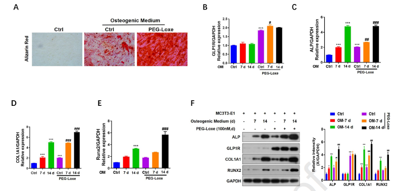
Fig. 1. Loxenatide enhances osteogenic differentiation and upregulates osteogenic markers via GLP-1 receptor activation.
A. Alizarin Red S staining for osteogenic changes. B–E. qPCR detection of GLP-1R, ALP, Col-1, and Runx-2 mRNA in osteoblasts. F. Western blotting of GLP-1R, ALP, Col-1, and Runx-2 protein levels in osteoblasts ( p < 0.05; p < 0.01; p < 0.001, # p < 0.05; ## p < 0.01; ### p < 0.001).
In vitro: Loxenatide inhibits osteoclast formation and inflammatory signaling pathways
The study used RANKL-stimulated RAW264.7 cells to induce osteoclast differentiation. Results showed that after loxenatide treatment, the staining intensity of osteoclast markers was significantly attenuated. Further molecular testing revealed that loxenatide reduced the mRNA expression levels of high mobility group box 1 (HMGB-1), tumor necrosis factor-α (TNF-α), interleukin-1β (IL-1β), and interleukin-6 (IL-6), and inhibited the expression of receptor for advanced glycation end products (RAGE), Toll-like receptor 4 (TLR4), and phosphorylated nuclear factor κB (NF-κB) proteins (Fig. 2). These findings suggest that loxenatide inhibits osteoclast differentiation and inflammatory responses through the HMGB1/RAGE/TLR4/NF-κB signaling pathway.
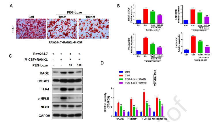
Fig. 2. Loxenatide inhibits osteoclast formation and inflammatory signaling pathways in osteoclasts.
A. TRAP staining for osteoclast bone resorption (100 nM loxenatide showed the most significant bone resorption effect). B. qPCR detection of HMGB1, TNF-α, IL-1β, and IL-6 mRNA in osteoclasts. C, D. Western blotting of RAGE, HMGB-1, TLR-4, pNF-κB, and NF-κB proteins in osteoclasts ( p < 0.05; p < 0.01; p < 0.001, # p < 0.05; ## p < 0.01; ### p < 0.001).
In vitro: Loxenatide inhibits osteoclast activity and inflammation by regulating the HMGB1 pathway
The study constructed an HMGB1 overexpression plasmid and transfected it into RAW264.7 cells. Results showed that HMGB1 overexpression partially offset the inhibitory effect of loxenatide on osteoclast differentiation and reversed the downregulation by loxenatide of inflammatory cytokines (TNF-α, IL-1β, and IL-6) and related signaling proteins (RAGE, HMGB1, TLR-4, and p-NFκB/NFκB) (Fig. 3). This further confirms that the bone-protective effect of loxenatide is primarily achieved through regulation of the HMGB1 signaling pathway.
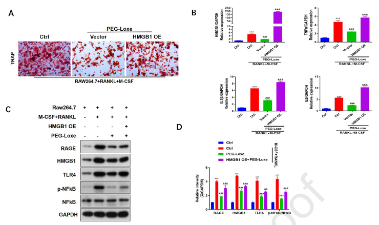
Fig. 3. Loxenatide inhibits osteoclast differentiation and inflammatory signaling pathways.
A. TRAP staining for bone resorption. B. qPCR detection of the effects of 100 nM loxenatide on HMGB1, TNF-α, IL-1β, and IL-6 mRNA in osteoclasts overexpressing HMGB1 plasmid. C, D. Western blotting of RAGE, HMGB1, TLR-4, and p-NF-κB/NF-κB proteins in osteoclasts overexpressing HMGB1 plasmid after 100 nM loxenatide treatment ( p < 0.05; p < 0.01; p < 0.001, # p < 0.05; ## p < 0.01; ### p < 0.001).
Animal study: Loxenatide ameliorates osteoporosis and increases bone mass in diabetic mice
The study used ApoE knockout mice fed a high-fat diet combined with streptozotocin to induce a T2DM model. After loxenatide intervention, serum levels of RAGE, HMGB1, TNF-α, and IL-1β were significantly reduced. HE staining and TRAP staining of femoral tissue showed reduced bone porosity and decreased osteoclast numbers in the loxenatide group. Micro-CT three-dimensional reconstruction analysis revealed significant improvements in trabecular bone mineral density, bone volume fraction, and trabecular thickness, along with increased cortical bone thickness and reduced porosity in the loxenatide group (Fig. 4). These findings indicate that loxenatide can improve bone microarchitecture and increase bone mass in diabetic mice.
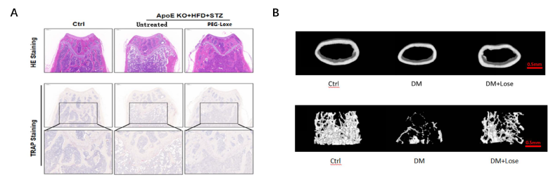
Fig. 4. HE staining, TRAP staining, and 3D micro-CT images of mouse femurs.
A. HE and TRAP staining for morphological changes in mouse femoral tissue. B. 3D micro-CT images of mouse femurs.
Clinical trial: Loxenatide effectively improves bone turnover status in T2DM patients with osteoporosis
The study enrolled 100 T2DM patients with osteoporosis and randomly assigned them to a loxenatide group or a non-loxenatide group, with a 52-week observation period. Results showed that levels of the bone resorption marker C-terminal telopeptide of type I collagen (CTX), as well as serum HMGB1, TNF-α, and IL-1β, were significantly lower in the loxenatide group than in the non-loxenatide group (p < 0.05). No significant difference in bone mineral density was observed between the two groups during the 52-week observation period. It should be noted that changes in bone mineral density typically require 18 to 24 months of continuous treatment to show significant differences, whereas bone turnover markers can change within 6 to 12 months after drug intervention and can serve as early indicators for monitoring drug efficacy. In terms of safety, no patients withdrew from the study due to gastrointestinal reactions or severe hypoglycemia.
Discussion and Summary
Through in vitro cellular experiments, animal models, and clinical research data, the investigators confirmed the bone-protective potential of loxenatide, mainly reflected in the following aspects:
- Promoting osteoblast differentiation and maturation
- Inhibiting osteoclast formation via the HMGB-1/RAGE/TLR4 signaling pathway
- Reducing inflammatory markers and improving femoral microarchitecture
- Improving bone turnover, reducing bone resorption, and directly promoting bone formation
The effect of GLP-1 RAs on bone metabolism is related to diabetes stratification and the degree of weight loss; using a low maintenance dose can preserve bone benefits. In this study, GLP-1 RA did not exacerbate bone loss in T2DM patients, possibly for the following reasons:
- GLP-1 RA directly promotes osteoblast activity and inhibits osteoclastogenesis
- Reduces the accumulation of advanced glycation end products (AGEs), decreases non-enzymatic glycation of bone matrix collagen, and reduces bone fragility
- Reduces inflammation and oxidative stress through multiple pathways, improving bone resorption
- Glycemic benefits partially offset bone loss associated with weight reduction
Among currently available GLP-1 RAs, loxenatide has a moderate effect on body weight intervention. The mean baseline body mass index (BMI) of patients in this study was 26.0 kg/m², which falls within the overweight range but does not meet the criteria for obesity. At the end of the trial, BMI decreased to approximately 25 kg/m², indicating limited weight loss. This may be one reason why loxenatide maintains bone benefits.
In summary, the study demonstrates that loxenatide can promote bone formation while inhibiting bone resorption through regulation of the HMGB1/RAGE/TLR4/NF-κB signaling pathway, exerting comprehensive bone-protective potential in T2DM patients with osteoporosis. This study is the first to validate the mechanisms of GLP-1 RA bone-protective effects at both the osteoblast and osteoclast levels, and fills the research gap on loxenatide in bone metabolism. Future multicenter, large-sample, long-term follow-up cohort studies will be conducted to evaluate the impact of loxenatide on fracture risk in T2DM patients.
References:
[1] Murray CE, Coleman CM. Impact of Diabetes Mellitus on Bone Health. Int J Mol Sci. 2019 Sep 30;20(19):4873.
[2] Lizhi Zhang, Jingjing Hu, Chunxiao Huang, et al. Effects of the glucagon-like peptide-1 receptor agonist PEG-Loxe on diabetic osteoporosis: a mechanistic study. Molecular Metabolism. Available online 1 June 2026, 102390. In Press. https://doi.org/10.1016/j.molmet.2026.102390

