


Over the past three to four decades, interventional cardiology has moved from spreading a technique to tackling complex lesions, and intravascular imaging, including intravascular ultrasound (IVUS) and optical coherence tomography (OCT), has grown from a research tool into a routine instrument of the catheterization laboratory. The supporting evidence for imaging guidance has become solid, with randomised trials such as IVUS-XPL, ILLUMIEN IV and ULTIMATE, together with multiple meta-analyses, consistently showing better outcomes. Real-world use rates and the quality of use, however, have not kept pace with that evidence. Where does the gap come from, and how can it be closed?
In October 2026, during the 37th Great Wall International Congress of Cardiology and Asian Heart Society Congress 2026 (GW-ICC/AHS 2026) in Beijing, the MedJ Cardiovascular Channel and GW-ICC launched a Great Wall Dialogue session on intervention and transformation, from technique dissemination to precision. Prof. Jianqiang Li of the First Affiliated Hospital of Harbin Medical University chaired the session. Prof. Gary S. Mintz, senior medical advisor of the Cardiovascular Research Foundation (CRF) and director of Transcatheter Cardiovascular Therapeutics (TCT), and Prof. Yang Cao of the same hospital were invited to speak. Prof. Mintz has witnessed intravascular imaging move from the laboratory into the catheterization laboratory, and Prof. Cao has worked on its clinical application for years. Their discussion of how the evidence should be read, how it can be put into practice, and what precision PCI means offers a clear line of reasoning for the questions above.
Article Key Points
• Evidence for imaging guidance is solid, but catheterization laboratory use rates still lag behind that evidence
• Prof. Mintz attributed the gap to three levels: cost, awareness and workflow, in ascending order of importance
• Imaging arms in IVUS-CHIP and OPTIMAL ran about 30 minutes longer, but the difference reflects workflow maturity
• Prof. Cao argued that the main obstacle is cognitive inertia, not a shortage of evidence
• OPTIMAL and IVUS-CHIP were neutral, yet a JACC meta-analysis still favoured imaging on outcomes including mortality
• A European Heart Journal analysis found larger benefit in East Asian trials, which Prof. Mintz linked to training
• The AHS position statement covers diagnosis and classification of calcified nodules, but device selection is unsettled
• Prof. Mintz called eruptive calcified nodules a nightmare, with recurrence of 30% to 50% and no solution yet
• Three rules carry the outcome: full stent expansion, no geographic miss, and avoidance of procedural complications
Expert Profile: Prof. Gary S. Mintz
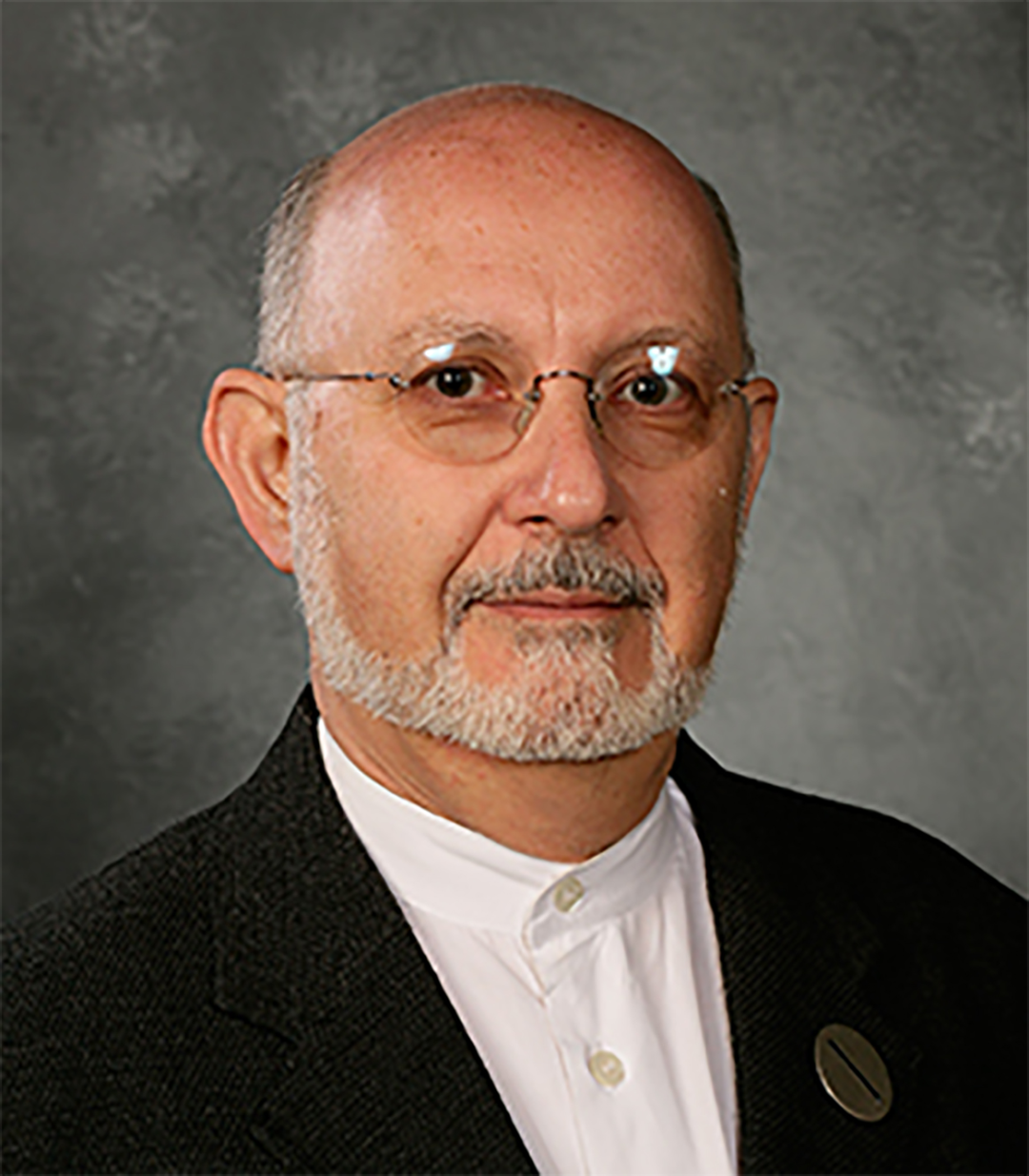
• Senior medical advisor of the Cardiovascular Research Foundation (CRF) and director of Transcatheter Cardiovascular Therapeutics (TCT)
• International authority on intravascular imaging; identified calcified nodules in the right coronary artery with IVUS in the 1990s
• Long-standing work on interpretation standards for intravascular imaging and on PCI optimisation; a major contributor to the Asian Heart Society (AHS) position statement on the management of coronary calcified nodules
Expert Profile: Prof. Yang Cao
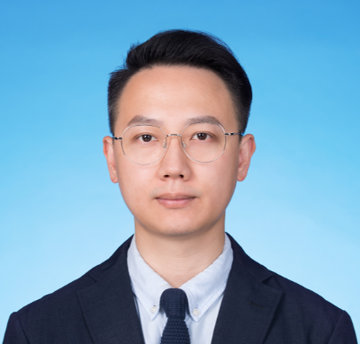
• Interventional cardiologist, Department of Cardiology, First Affiliated Hospital of Harbin Medical University
• A well-known Chinese expert in interventional cardiology, with extensive experience in the clinical application of intravascular imaging
• Visiting fellow at the CRF ten years ago, where he trained under Prof. Mintz; contributor to the AHS position statement on the management of calcified nodules
Expert Profile: Prof. Jianqiang Li
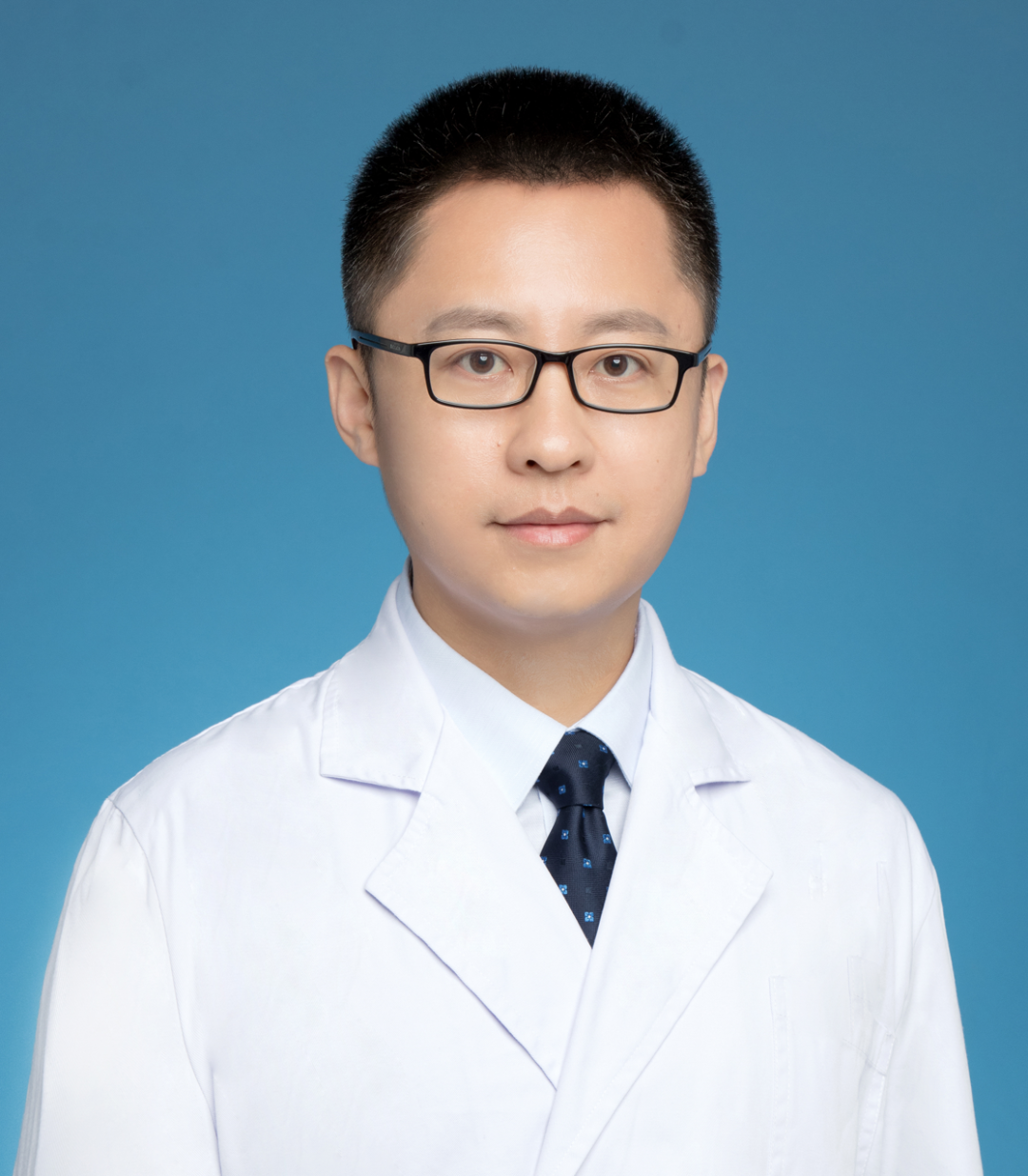
• Chief physician, professor and master’s supervisor, First Affiliated Hospital of Harbin Medical University
• Member of the Cardiovascular Clinical Research Group, Chinese Society of Cardiology
• Member of the Cardiovascular Metabolism Medicine Panel, National Cardiovascular Disease Expert Committee
• Standing committee member of the Integrated Oncology Cardiology Branch, Chinese Anti-Cancer Association
• Member of the Hypertension Branch, Chinese Geriatrics Society
Three Sources of the Gap: Cost, Awareness, and Workflow
Why is the evidence sufficient while use remains insufficient? Prof. Mintz reduced the reasons to three levels, in ascending order of importance.
The first is economics. In some less developed regions, an intravascular imaging catheter costs around 1,000 US dollars, with no insurance coverage and the cost borne by the patient, so price becomes a practical barrier that cannot be avoided. By comparison, catheter prices in China have fallen to several hundred US dollars and the economic threshold is falling; over a wider range of settings, however, price remains the first constraint.
The second is awareness, and this is the emphasis of Prof. Mintz. Education here does not simply mean learning to read images. It is a layered structure: understanding what the images mean, mastering how that information is converted into clinical decisions, and, most importantly, recognising what the best result of PCI looks like and being willing to work for it, rather than settling for a result that is good enough.
The third is workflow, which shows up chiefly as concern about procedural time. In IVUS-CHIP and OPTIMAL, imaging arms took about 30 minutes longer than angiographic arms, and this is often read as evidence that imaging slows procedures down. Prof. Mintz pointed out that the difference is not inherent to the technology but reflects how mature the workflow is: in the Columbia University-led centre where he works, imaging systems are kept ready, imaging guidance has become routine, and three imaging acquisitions take only five minutes. Most of the extra time therefore comes from inadequate training and from the absence of workflow integration.
The Last Mile: From Guideline Recommendation to the Operating Table
The evidence and the guidelines are in place; what remains is to turn recommendations into the habits of each individual operator. Prof. Cao has a first-hand view of how long that road is.
His training at the CRF ten years ago allowed him to compare the cognitive differences between operators in China and the United States at close range. He stated plainly that the main obstacle to implementation is not a shortage of evidence but cognitive inertia: operators who have worked with angiography for decades tend to assume that a good angiographic result is equivalent to a good intravascular result, and so undervalue the incremental value of imaging.
He proposed that the value of intravascular imaging should be understood as a complete clinical chain rather than an isolated act of image acquisition. Specifically: at the reading stage, identifying true bifurcations and left main lesions; at the strategy stage, deciding on a single-stent or a two-stent approach from the imaging findings and adjusting dynamically during the procedure; and at the finishing stage, optimising the stent and carrying the information into follow-up. The three stages are interdependent, and any missing link reduces the value of imaging. What keeps the chain running reliably in daily practice, he argued, is a mature, executable flowchart.
Notably, this judgement is not shaken by individual neutral studies, and how to read those neutral studies rationally was the next question in the conversation.
Reading the Neutral Trials: OPTIMAL and IVUS-CHIP Revisited
Neither IVUS-CHIP nor OPTIMAL produced a positive result, and at one point this raised doubts about the value of imaging.
Take OPTIMAL as an example. Its optimisation targets followed the consensus values from 2011: a minimum stent area of 5 mm², 6 mm² at the circumflex ostium, 7 mm² in the left anterior descending artery, 7 mm² at the polygon of confluence and 8 mm² in the left main. The problem, he said, is that this standard was originally derived to predict 9-month angiographic restenosis rather than clinical events. A recent single-centre study re-derived area thresholds from 5-year clinical outcomes and obtained clearly larger values, and the old thresholds applied retrospectively to 5-year outcomes did not predict clinical events. On that basis Prof. Mintz judged that the study paired the wrong standard with the wrong outcome. In addition, there has been no core laboratory analysis, so it is not possible to confirm that the targets were met. The proportion of cases in which the targets were achieved in IVUS-CHIP is also in question, with recent data suggesting it may have been as low as 50%. Prof. Cao added that the proportion of patients in whom IVUS was actually used in both trials, roughly 60% before the procedure and 90% in total, was lower than in the positive studies.
Placed back into the whole pool of evidence, the conclusion becomes clearer. A meta-analysis in the Journal of the American College of Cardiology showed that even with the two neutral trials included, outcomes in the imaging group, including mortality, remained better. An analysis in the European Heart Journal found greater benefit in East Asian trials, which Prof. Mintz attributed to better training. As he put it, of roughly 300 relevant studies, only three were negative. The two neutral trials therefore do not overturn the overall direction of the evidence; they only underline that method and standards matter just as much.
Calcification and Calcified Nodules: Tool Limits and Lesion Risk
Discussions of method and standards eventually come back to how a specific lesion is read on imaging. Calcification is the lesion type that tests the limits of a tool first, and that exposes the gap most clearly between having a tool and knowing how to use it. OCT can measure calcium thickness and calculate area and volume, whereas IVUS cannot penetrate calcium and can only measure the arc and the length of the calcified segment. Prof. Mintz pointed out that with IVUS the arc and length must be used fully, whereas with OCT the blind spots must be watched for: if there is lipid plaque on the surface of the calcium, deep calcium may be completely invisible.
For ordinary calcification the tools each have their boundaries, and the real gap lies with the user. Within the calcification spectrum, however, calcified nodules are more difficult than ordinary calcium and place a greater demand on imaging interpretation and clinical judgement. They are uncommon, but they are one of the triggers of acute coronary syndrome (ACS); more importantly, even when stent optimisation is completed in line with guidelines, target lesion failure remains concentrated in this lesion type. Recognition is the first hurdle, because angiography cannot display a nodule and its appearance can resemble the filling defects of thrombus or dissection.
Prof. Mintz identified this lesion with IVUS in the right coronary artery as early as the 1990s, but missed diagnoses remain common even now. Precisely because recognition is difficult, classification becomes a precondition for judging prognosis: eruptive and non-eruptive nodules behave very differently, and OCT is better at telling the two apart. This year the Asian Heart Society (AHS) published a position statement on the management of coronary calcified nodules, establishing a framework for diagnosis and classification, while the treatment side remains blank. How to select devices, and whether drug-eluting stents (DES) or drug-coated balloons (DCB) are preferable, are both unresolved for want of large randomised studies; Prof. Cao said his team is preparing a clinical trial to follow up on this.
While treatment options remain unsettled, Prof. Mintz narrowed the concept of a calcified nodule itself. He pointed out that today any small protrusion on calcium is called a calcified nodule, and that some papers put the figure at 40% or 60% of lesions, which in his view is unfounded. Non-eruptive nodular calcium resembles a calcified boulder covered by an intact fibrous cap; it is difficult to treat in the acute phase and may require lesion modification with intravascular lithotripsy (IVL), rotational atherectomy or orbital atherectomy, yet the long-term prognosis is not poor. The truly dangerous lesion is the eruptive calcified nodule, which is the third most common cause of ACS and in the acute phase needs only a stent, yet has a recurrence rate of 30% to 50%, a rate of recurrence beyond the second episode above 50%, and intervals between episodes that keep shortening. He called this a nightmare and admitted that no solution is yet available. And if there is no ideal device, the only rational choice is restraint: do not intervene on a lesion without haemodynamic significance.
Separating Seeing from Measuring: Three Rules for Precision PCI
Restraint is not the abandonment of judgement but the handing of judgement to the more appropriate method. Imaging answers what can be seen clearly, physiology answers whether the lesion should be treated, and Prof. Mintz allocated the two by location. For left main lesions, imaging comes first, because the long-term data are far more extensive than for physiology and the quality of treatment is decisive. For non-left main lesions the reverse applies: some 30 to 40 studies show that the positive predictive value of imaging for functional significance is only around 50%, whereas physiological assessment has greater predictive value and is supported by large randomised trials, and the pressure pullback gradient can distinguish focal from diffuse disease. Prof. Mintz also calibrated one detail: a post-stent fractional flow reserve (FFR) of 0.9 is generally regarded as optimal, but for the left main, values of 0.80 to 0.84 are acceptable. Prof. Cao added that physiology answers whether to intervene at this moment, while future events also depend on smoking, lipids, diabetes and plaque vulnerability, and that a vulnerable plaque does not automatically mean a stent should be implanted, since drug therapy may shrink it and stabilise it.
The timing of judgement can also be moved earlier. The CorVIS system, developed by the team of Prof. Yue Li at the First Affiliated Hospital of Harbin Medical University, takes coronary computed tomography angiography (CTA) as its input and uses artificial intelligence (AI) to perform three-dimensional reconstruction, so that left main disease, severe calcification and chronic total occlusion (CTO) can be mapped before the procedure, showing where the calcium lies, whether it is circumferential, and the distal state and entry orientation of the CTO. Prof. Mintz was both welcoming and cautious about this. He said that AI iterates extremely quickly, so that a judgement made today may be out of date tomorrow, and that the algorithms and thresholds will need to be recalibrated and revalidated; both the potential and the need for continuous verification should be kept in view.
However the judgement is made, before or during the procedure, it returns to the technical execution. Standardised medical therapy is the foundation, and on that foundation the long-term outcome is decided by the procedure. Prof. Mintz reduced this to three principles that must be followed, in a fixed order.
First, the stent must be fully expanded.
Second, the stent edges must avoid the lesion, so as not to suffer geographic miss.
Third, procedural complications must be avoided.
These three principles are not craft that depends on individual experience but procedural steps that can be fixed through training and workflow, which is why promoting them and pursuing precision are not in conflict. Prof. Cao pointed out that standardisation and individualisation are in fact the same story: imaging use in China is rising, but its distribution across regions is uneven, and the priority should be systematic training rather than equipment purchase alone.
Closing
The final part of the conversation turned to young doctors about to enter the field. Prof. Cao shared two lessons from the past decade: first, that learning must not stop, which he regarded as the most valuable gain from his time as a visiting fellow at the CRF; and second, that the patient always sits at the centre of the operation, not the device, not the tool and not the image. Prof. Mintz’s advice focused on how to choose an environment for training: an institution with a sufficient volume of cases, good teachers and complete technical and device coverage, so that, as he put it, by the time training is finished you can reach the best level possible.
Interventional cardiology has reached a point where having done the procedure is no longer the question; doing it correctly is the real divide. The value of intravascular imaging does not sit inside the device. It sits in the hands of the people who use it.

