


A 45-year-old Ms. Zhang, born with a double uterus and double vagina, had undergone three cesarean deliveries over the past decade before finally receiving definitive surgical treatment recently.
In an interview with MedJ, Prof. Jian Cheng, director of Gynecology Ward II at Xi'an Central Hospital, reviewed the full course of this high-difficulty operation.
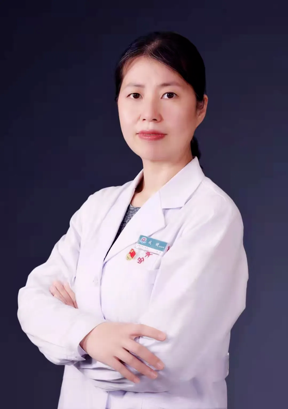
Fig. 2. Prof. Jian Cheng from Xi'an Central Hospital.
An Anatomy That Defied Expectations
Ms. Zhang had long suffered from heavy and prolonged menstrual bleeding as well as dysmenorrhea. Persistent dyspareunia and abdominal pain affected her quality of life, while enlarging uterine fibroids worsened chronic blood loss and progressively aggravated her anemia.
She had previously undergone three cesarean sections, an appendectomy and bilateral tubal ligation, leaving her with a complex history of open abdominal surgery. Only when she recently presented to the gynecology department at Xi'an Central Hospital did she fully learn how unusual her condition was.
"She had previously been diagnosed with a vaginal septum, meaning a wall inside the vagina partially or completely dividing it into two channels. But after more detailed evaluation, we found a more complex reproductive tract malformation, with duplicated structures extending from the vagina to the uterus. Each uterus was connected to its own ovary and fallopian tube, and even the ureters were displaced," Prof. Cheng explained.
This time, Ms. Zhang hoped a minimally invasive procedure could finally resolve the gynecologic problems that had troubled her for years. She had sought care at multiple local hospitals, all of which advised referral to a higher-level center with extensive experience.
Comprehensive assessment showed that this would be an exceptionally difficult minimally invasive gynecologic operation. The greatest challenge was the extreme anatomic abnormality: with a double uterus, double cervix and double vagina, the operative field available for localization and instrument handling was very limited.
Multiple prior laparotomies had also caused extensive, dense pelvic adhesions, completely obscuring the normal layers of anatomy. Any misstep during surgery could have injured vital organs such as the ureters, bladder or bowel.
"The abnormal anatomy had an even greater impact on the procedure. It completely broke with routine anatomic expectations, so we could not operate by referencing a normal body structure. Every step required precise judgment," Prof. Cheng told MedJ.
More importantly, the team needed to complete laparoscopic resection of both uteri and both fallopian tubes, meticulous adhesiolysis for severe pelvic adhesions, and resection and correction of the vaginal septum during the same operation. That demanded highly advanced endoscopic technique, a strong command of pelvic anatomy and rapid intraoperative decision-making.
Oncologic Surgery Experience as a Safety Net
To overcome these difficulties, Prof. Cheng and colleagues carried out extensive preoperative planning. They completed imaging-based reconstruction of the anatomic relationships, anticipated the sites and trajectories of adhesions, prepared contingency plans for high-risk injuries involving the ureters, bladder and bowel, and fixed the operative sequence in advance: first restore the anatomy through adhesiolysis, then remove the lesions and correct the malformation.
The operation lasted more than three hours—more than triple the usual time for similar surgery. At the start, the surgeons could not even visualize the uterus under laparoscopy because the bowel and greater omentum had completely covered the reproductive organs. Using an ultrasonic scalpel and a combination of sharp and blunt dissection, the team gradually separated the omentum and bowel, even accepting limited sacrifice of the uterine serosa to avoid damaging the bowel or bladder.
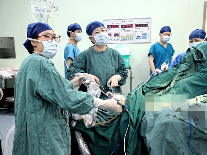
Fig. 3. Intraoperative image from the source article.
Abnormal pelvic anatomy posed another major challenge. Preoperative MRI had not clearly shown the exact relationship between the cervix and bladder. Once inside the abdomen, the surgeons found that the patient's two cervices were tightly apposed, while the bladder had also been reshaped by the duplicated uterus, with part of the bladder tissue extending between the two uterine bodies.
Because the reproductive and urinary systems share common embryologic origins, an abnormality in one system may be accompanied by changes in the other. The broad uterine mass directly altered the course of both ureters. According to the article, the literature currently lacks domestic and international guidelines on surgical techniques for patients with uterine malformations, and minimally invasive procedures may need conversion to open surgery because of anatomic variation.
There were also smaller intraoperative challenges. Standard instruments were designed for a single cervix, so the team repeatedly verified whether the left cervix had been completely removed. "Before and after dealing with the vaginal septum, we carefully rechecked the cervical ends and only felt reassured once we confirmed that both had been fully excised with no residual tissue," Prof. Cheng said.
She noted that the department mainly focuses on gynecologic oncology and pelvic floor surgery, disciplines that require precise dissection of pelvic and abdominal vessels, lymphatics and nerves. Experience gained from cervical cancer surgery had trained the team to separate the ureters and bladder safely, while the variety of pelvic floor procedures had made them highly familiar with the anatomy and risk points around the vagina and cervix.
Not long ago, the department also completed a difficult operation for a uterine accessory cavity with highly atypical preoperative findings and imaging features. This rare obstructive reproductive tract malformation has been reported in only a little more than 100 cases worldwide.
The surgery for Ms. Zhang was ultimately successful. The team completely removed the lesions and finished correction of the vaginal septum. She recovered smoothly after the operation, experienced only mild pain, got out of bed early and rapidly regained gastrointestinal function. The dyspareunia, abnormal uterine bleeding and anemia that had troubled her for more than a decade were fundamentally resolved.
Reflecting on the case, Prof. Cheng said the team also learned from the procedure: when managing broad and complex adhesions, combined sharp-and-blunt dissection works well, and clearly identifying the distribution of major organs and vessels before starting at a safe point can markedly reduce the risks of bleeding and organ injury.

