


Recently, The BMJ (Impact Factor = 93.6) published an article from the Neurofibromatosis (NF) team at Shanghai Ninth People's Hospital, affiliated with Shanghai Jiao Tong University School of Medicine. The article reported an extremely rare clinical case.
The first author of the paper, Dr. Liu Jun, is a postgraduate student in the eight-year clinical medicine program at Shanghai Jiao Tong University School of Medicine and a member of the "post-00s" generation. Under the guidance of Professor Li Qingfeng and Associate Professor Wang Zhichao, she spent over six months writing and revising the manuscript, which was successfully published in The BMJ on December 12 of last year.
What was so rare about the case that it made it to one of the top four medical journals? How did she discover this rare case, and what challenges did she face during the submission process? Dr. Liu Jun shared the detailed process with "MedJ."
The BMJ published an article by the Neurofibromatosis team at Shanghai Ninth People's Hospital. The first author is Dr. Liu Jun, and the corresponding author is Associate Professor Wang Zhichao. / Screenshot from The BMJ
Surprising Test Results
The case published in The BMJ involved a rare presentation of Type 1 neurofibromatosis (NF1) with intrathoracic meningocele.
Type 1 neurofibromatosis is a disease caused by mutations in the NF1 gene. Patients typically present with multiple subcutaneous tumors all over the body, resembling bubbles on the skin. These "bubbles" are essentially neurofibromas. They can also grow in internal organs, the spine, and the brain, causing multisystem complications. In severe cases, patients may develop deformities or paralysis, and there is a risk of malignancy that can be life-threatening.
The patient, Ms. X, is a 53-year-old woman with NF1 who visited the NF specialty clinic at Shanghai Ninth People's Hospital at the end of 2023. "The patient walked in by herself, accompanied by her daughter. Although she was not very talkative, her mental state was still acceptable, and she did not show obvious discomfort," Dr. Liu Jun said. She and Associate Professor Wang Zhichao jointly received Ms. X. The patient had a huge plexiform neurofibroma on her face, and their initial reaction was that she wanted to have the facial tumor removed.

Photo provided by Dr. Liu Jun
However, after listening to the patient's daughter describe the condition, they realized that Ms. X's situation was more complicated than they had anticipated.
According to the daughter, Ms. X was diagnosed with NF1 at an early age. In 2017, she began to experience coughing and shortness of breath, but since these symptoms did not significantly affect her life, she did not pay much attention to them. About a year before this visit, her breathing difficulties worsened significantly. She had to stop and rest after walking a short distance, so she went to a local hospital for a check-up.
The local hospital arranged a CT scan for Ms. X. The results were shocking to both the patient and her family: her right thoracic cavity was completely occupied by a huge cystic lesion, like a giant stone pressing on a balloon, causing the right lung to almost completely collapse. The trachea and mediastinum, which should have been in the center of the chest, were pushed to the left side. Additionally, there was a smaller mass in the left thoracic cavity.
What were these masses? After visiting several local hospitals, Ms. X was told that they might be neurofibromas, malignant tumors with secondary effusion, or meningoceles. However, no doctor could provide a definitive diagnosis.
Research has shown that patients with NF1 have a higher risk of developing malignant tumors compared to the general population. If the masses were malignant tumors, treatment would need to be initiated as soon as possible to prevent tumor progression and metastasis.
However, without a clear diagnosis, it was impossible to determine the next treatment plan. Ms. X and her family were left in a state of worry.
How to Make a Definitive Diagnosis?
The same question also troubled Dr. Liu Jun and Associate Professor Wang Zhichao, as well as the senior medical editors at The BMJ. During the manuscript revision process, the editorial team had asked: How can we definitively diagnose the huge mass as a meningocele?
"The meninges surround the surface of the spinal cord. Normally, they do not protrude because of the bony protection of the spine. However, Ms. X had a thoracic vertebral defect due to NF1, like a missing brick in a wall, allowing the meninges to protrude through the gap and form a small pocket. Cerebrospinal fluid accumulated in the pocket, increasing over time and eventually filling the entire right thoracic cavity," Dr. Liu Jun explained.
In fact, the NF specialty clinic had previously seen a few patients with NF1 and intrathoracic meningoceles, but none had such a large mass. Dr. Liu Jun told "Medical Community" that since NF1 with meningoceles is extremely rare, it is essential to differentiate it from neurofibromas in patients with NF1 who have masses.
Last December, another tertiary hospital in China also received a female patient with dyspnea and a large mass in the thoracic cavity. The doctors used a thoracoscopic-assisted incision to remove a 7-jin (about 3.5 kg) tumor from her body, and the pathological result was a neurofibroma.
It is important to note that the treatment plans for intrathoracic meningoceles and neurofibromas are completely different. Using thoracoscopy without distinguishing between the two could lead to severe consequences.
"If it is a meningocele, the thoracoscopic instruments would puncture the cyst as soon as they are inserted, filling the entire thoracic cavity with cerebrospinal fluid, making the situation uncontrollable," Dr. Liu Jun said. Misdiagnosing a meningocele as a neurofibroma could also lead doctors to prescribe NF1-targeted drugs, subjecting patients to expensive and inappropriate treatments.
To make a definitive diagnosis, the department applied for a magnetic resonance imaging (MRI) scan for the patient based on the previous examinations. The MRI showed that both masses had uniform high signals on T2-weighted sequences, consistent with cerebrospinal fluid, rather than the heterogeneous slightly high signals typically seen in neurofibromas.
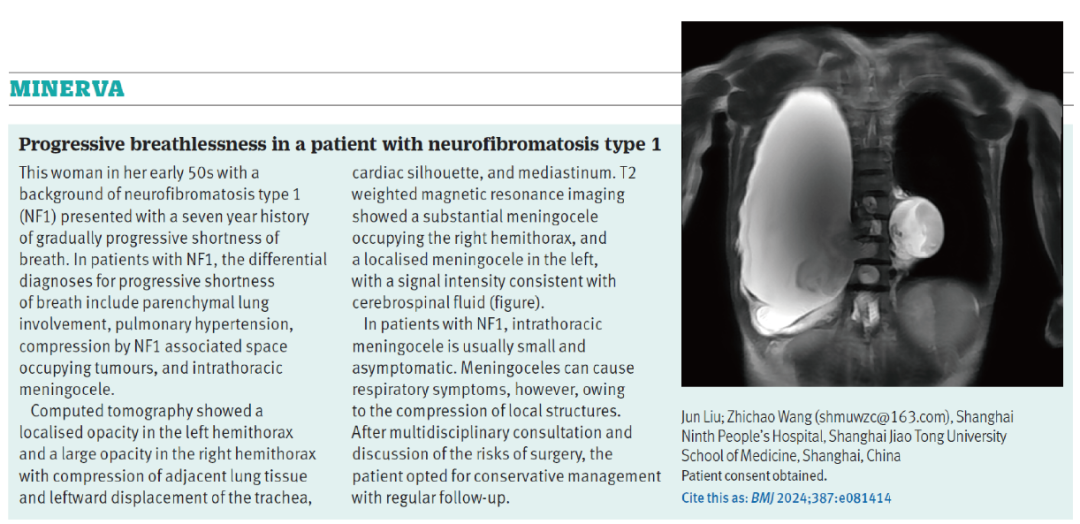
The patient's chest MRI shows that the right thoracic cavity is completely occupied by a cystic lesion. / Screenshot from The BMJ
The NF treatment team at Shanghai Ninth People's Hospital, together with experts from thoracic surgery, radiology, neurosurgery, and spinal surgery, held a multidisciplinary consultation. Based on the patient's medical history, clinical manifestations, imaging results, and laboratory tests, the experts diagnosed both masses as meningoceles.
Regarding the treatment of this patient, the experts who participated in the multidisciplinary discussion believed that a multidisciplinary joint surgical approach could be adopted, with the best option being a large-incision thoracotomy for drainage and repair. In layman's terms, this means tying off one end of the cyst to block the "inlet," releasing the cerebrospinal fluid through a drainage tube, and then performing the repair.
Given the large size of the mass in the patient's thoracic cavity, to avoid complications such as atelectasis and pulmonary edema due to sudden relief of lung pressure, as well as complications from low intracranial pressure caused by massive loss of cerebrospinal fluid, the patient might need multiple surgeries. This also posed higher demands on perioperative care and corresponding treatments.
Overall, while the surgical treatment plan could resolve the issue, it was time-consuming and high-risk. After the experts informed the patient and her family of the surgical plan and risks, the patient and her family, after weighing the surgical risks against the impact on their current quality of life, ultimately chose a conservative treatment plan.
Currently, the patient is undergoing regular follow-ups and close monitoring. If her breathing difficulties continue to worsen or if she develops other new respiratory symptoms, she will need to return to the hospital for re-evaluation and to reassess whether surgery is necessary.
Rejected by NEJM
After Ms. X left, the NF treatment team at Shanghai Ninth People's Hospital systematically organized the patient's information and reviewed the case characteristics. During the discussion, Associate Professor Wang Zhichao keenly identified the rarity and uniqueness of the case and guided Dr. Liu Jun to delve deeper into the mechanisms behind this type of case and to search for any relevant literature reports.
"After systematically reviewing the literature on respiratory manifestations in NF1, we found that this patient's presentation was highly atypical and worth reporting," Dr. Liu Jun told "MedJ."
Many top-tier medical journals have case report sections. Compared to articles on basic research and clinical trials, case reports are often shorter and labor less-intensive, but many cases have clinical value worth exploring and sharing.
Publishing case reports in top-tier journals allows clinicians to exchange ideas with peers worldwide, leveraging the journals' significant medical influence. This is a challenge that many clinical doctors continuously attempt.
However, cases that make it to top-tier journals often have distinct features such as "extremely rare" or "highly interesting," which is why many doctors believe that publishing case reports in top journals is "a matter of luck."
How rare was Ms. X's case? Dr. Liu Jun explained that NF1 itself is a rare disease with a global incidence rate of about 1 in 2500.
Epidemiological data on NF1 is scarce, and finding literature on NF1 with respiratory distress is like searching for a needle in a haystack.
Dr. Liu Jun spent over three months collecting and reviewing literature before deducing that approximately 10% of all NF1 patients experience respiratory distress, and about 10% of those with respiratory distress have meningoceles. Meningoceles as severe as Ms. X's are even rarer in the published literature.
In May of last year, Dr. Liu Jun finished writing the article. She initially submitted it to The New England Journal of Medicine (NEJM) with high expectations but quickly received a rejection letter.
Despite feeling somewhat discouraged, Dr. Liu Jun did not give up. Under the guidance of Associate Professor Wang Zhichao, she reviewed influential medical journals that had published case reports in recent years and ultimately focused on another top-tier journal with a case report section—The BMJ.
In fact, shortly before Dr. Liu Jun's submission, The BMJ had published several cases focusing on cystic lesions, which had a similar presentation to Ms. X's case, giving her some confidence.
Two months after submitting to The BMJ, the editorial team sent an encouraging message: after consulting with many doctors, they also believed that the case was extremely rare and of significant clinical value, and they recommended it for publication.
However, the editorial team also provided Dr. Liu Jun with revision suggestions, asking her to supplement the detailed records of the multidisciplinary consultation and the patient's chest X-ray. Considering the greater diagnostic value of MRI, the department had not previously performed an X-ray on the patient.
When the revision suggestions were received, the patient had been discharged for several months. How could they supplement the examination?
Dr. Liu Jun explained that since 2010, the department had registered the clinical information of every NF1 patient admitted to the specialized disease database. After discussing with Associate Professor Wang Zhichao, they contacted the patient according to the standard procedure. With the understanding and support of the patient and her family, they arranged for the X-ray to be done at a local hospital.
In the next round of revisions, the The BMJ editorial team also asked Dr. Liu Jun to supplement the differential diagnosis methods for respiratory distress in NF1 patients. Dr. Liu Jun then conducted a systematic literature review and included a summary table of all NF1 respiratory distress-related literature in her response to the editors, rigorously discussing the need to differentiate NF1 respiratory distress from interstitial lung disease, tumor compression, and pulmonary hypertension.
At the same time, while reviewing the literature, Dr. Liu Jun found that compared to other imaging methods, MRI played an important role in differential diagnosis, which aligned with the clinical experience of the NF team at Shanghai Ninth People's Hospital. After communicating this finding with the editorial team, they decided to use the MRI images for the case presentation.
After nearly two months and four rounds of careful revisions, on November 18th of last year, the editorial team finally accepted the case report.
Publishing a case report in a top-tier journal is not just about luck
On December 12th of last year, the article was published online on the official website of The BMJ and appeared in print two days later.
This meant that Dr. Liu Jun, as the first author, had achieved an impact factor of over 93 points in a single article. Many people congratulated Dr. Liu Jun, but some also said that publishing a top-tier journal article based on a single case was just "good luck."
"Luck is a factor, but it is also based on an understanding of top-tier journals," Dr. Liu Jun told "Medical Community." After being rejected by NEJM, Associate Professor Wang Zhichao and she carefully analyzed the reasons and found that NEJM prefers to publish interesting cases and clinical presentation photos rather than diagnostic images of patients.
Therefore, when submitting to The BMJ, Dr. Liu Jun did more preparatory work. She read a large number of previous articles in the case report section, analyzed the characteristics of accepted cases, and learned how to use the language style of The BMJ case reports. She condensed the patient's more than seven-year medical history into just over 150 English words, and after repeated discussions and revisions with Associate Professor Wang Zhichao, she cautiously submitted the first version of the article.
In Associate Professor Wang Zhichao's view, Dr. Liu Jun's success in publishing a high-impact article was also inseparable from the nearly 60 years of in-depth work by the plastic surgery department of Shanghai Ninth People's Hospital in the field of neurofibromatosis.
In 1969, under the leadership of Academician Zhang Disheng, the plastic surgery department of Shanghai Ninth People's Hospital was the first in China to open a neurofibromatosis specialty clinic. With the strong support of Vice President and Head of Plastic Surgery, Professor Li Qingfeng, the neurofibromatosis diagnosis and treatment team established the Shanghai Ninth People's Hospital Neurofibromatosis Diagnosis and Treatment Center.
So far, the center has treated more than 9,000 patients and is one of the largest neurofibromatosis centers in China. It has also established the largest neurofibromatosis database in China and the second-largest in the world, as well as the largest clinical sample bank for neurofibromatosis globally.
"Only after seeing so many patients with neurofibromatosis can one recognize the clinical value of a special case. If one is not familiar enough with the disease, they can only suggest a referral and miss the opportunity to publish in a top-tier journal," said Associate Professor Wang Zhichao. Every patient that a doctor sees may hold a "golden nugget" of a valuable and clinically rich case.
"Chinese doctors are not short of case numbers; what they need more is the ability to 'spot gold,'" said Associate Professor Wang Zhichao.

