


Cardiovascular imaging provides the basis for diagnosis and treatment decisions, yet once the images have been acquired, a substantial amount of measurement and analysis still depends on the time of physicians. The development of artificial intelligence (AI) is changing this. Some quantitative imaging analyses that used to take hours can now be returned within minutes. When AI takes over part of the measurement and analysis, what happens to the work of imaging specialists? And what does a new technology have to go through before it moves from research into the hospital?
From October 15 to 18, 2026, the 37th Great Wall International Congress of Cardiology and Asian Heart Society Congress 2026 (GW-ICC/AHS 2026) was held in Beijing. The Great Wall Dialogue, the official branded column launched jointly by the MedJ Cardiovascular Channel and GW-ICC, presented a session titled “Imaging and Intelligent Transformation: From Imaging Gold Standards to AI-Enabled Precision Decision-Making”. It was chaired by Prof. Jiapeng Chu of the Second Affiliated Hospital of Soochow University, with Prof. Nico Bruining, head of digital cardiology at the Erasmus University Medical Center (Erasmus MC) in Rotterdam and founding editor-in-chief of European Heart Journal - Digital Health, as the guest. The conversation addressed how AI changes imaging analysis, how it moves into clinical practice, and how it can reach more patients.
Article Key Points
• AI now returns fully automated echocardiographic analysis in minutes instead of hours
• Imaging specialists are not replaced at present; their work shifts to reviewing AI output and integrating data
• External validation in other independent centres should precede prospective trials for AI in imaging
• Prof. Bruining prefers monitoring after deployment to post-market surveillance for clinical AI tools
• AI-guided imaging may extend basic cardiac checks into primary care and nurse practitioner services
Expert Profile: Prof. Nico Bruining
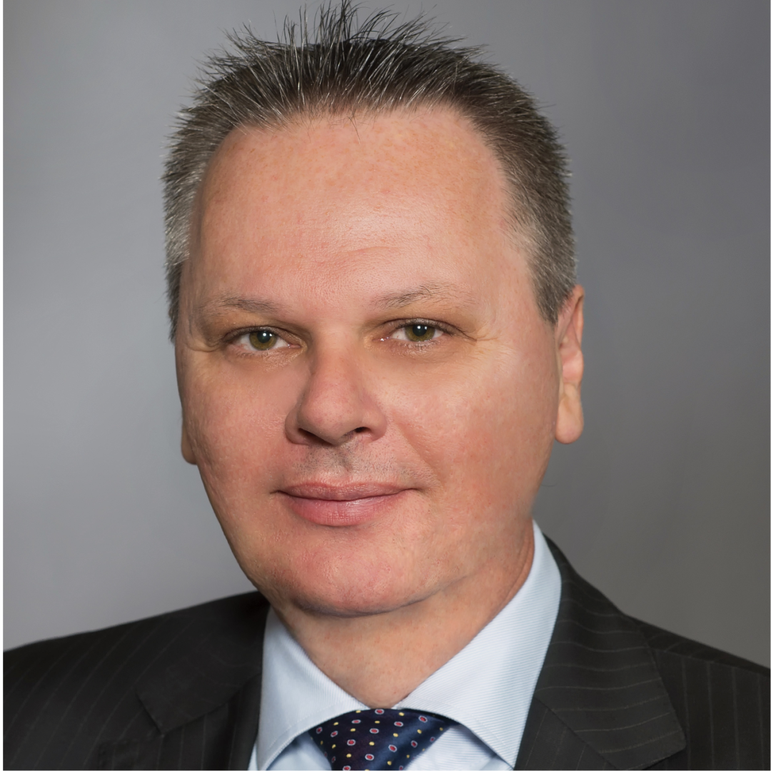
• Head of the Department of Digital Cardiology, Thorax Center, Department of Cardiology, Erasmus University Medical Center (Erasmus MC), Rotterdam, the Netherlands
• Founding editor-in-chief of European Heart Journal - Digital Health, the official digital health journal of the European Society of Cardiology (ESC)
• Within the ESC, leads the digital cardiology division of the Digital Cardiology and Artificial Intelligence (DCAI) Committee and co-chairs the scientific programme of the Digital Cardiology and AI Summit; member of the congress programme committee and its digital health programme
• Works in the catheterization laboratory alongside pioneers in interventional cardiology, Profs Patrick Serruys and Pim de Feyter; studied technical computer sciences and joined the Digital Cardiology Department as a scientist after graduating in 1993
• Research centred on cardiovascular imaging to evaluate new therapies; his methods and techniques have been used in several first-in-man interventional studies, including the development of 3D echocardiography
Expert Profile: Prof. Jiapeng Chu
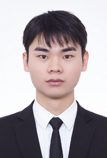
• Attending physician, Department of Cardiology, The Second Affiliated Hospital of Soochow University, Suzhou, China
• Doctor of medicine from Tongji University; visiting scholar at the National University of Singapore
• Long-standing work in clinical practice, teaching and research in cardiology, with experience in the diagnosis and treatment of hypertension and coronary heart disease
• Main research interests in cardiovascular imaging and in basic and translational research on hypertension
• Principal investigator on one municipal project in Suzhou and co-investigator on national and provincial programmes; more than 20 SCI-indexed papers, 10 as first author; reviewer for several cardiovascular and metabolic journals
Will AI Replace Imaging Specialists?
“AI is now a genuine accelerator,” said Prof. Bruining. Some computer-assisted quantitative analyses that previously took hours can now return fully automated echocardiographic analysis within minutes. In the catheterization laboratory, analyses that previously had to be completed offline after the examination can also be performed during the procedure, which shortens the waiting time of patients and reduces transfers.
So, as measurement and analysis are increasingly done by tools, will imaging specialists be replaced? Prof. Bruining’s judgement is that this is not the case at present, but that the content of their work is changing. Doctors will do more reviewing of AI-generated results, more integration of information from different sources, and more judgement in the light of the individual patient. With some repetitive operations removed, doctors can spend more time on understanding the patient and discussing the condition.
The transition, however, requires gradual adaptation by the team. In Prof. Bruining’s observation, younger doctors usually take up new technology readily, whereas some colleagues prefer to observe how it performs in practice; some nursing staff and ultrasound technicians remain concerned about the reliability of the tools and about the effect on their roles. For doctors, the requirement is not only to learn to use AI but also to understand how it works and to review its output critically.
From Research to Clinical Practice: What Evidence Does AI Need?
AI models with excellent performance keep emerging, but the applications that actually enter routine diagnostic workflows remain limited. On this gap between development and practice, Prof. Bruining said: “We need solid clinical evidence.” Many studies have already produced high-quality model development work; the next step is to take these tools out of the research environments in which they were developed and subject them to broader external testing.
His proposal is that, once a model has been developed, external validation should first be carried out in other independent centres, followed by prospective clinical trials. Research should demonstrate not only that a technique works, but also how applicable it is and how it performs in different health systems.
International collaboration is an important part of moving this forward. He noted that technology developed in China should be validated in other health systems such as Europe, and that technology developed in Europe needs validation in other regions such as China and the United States. Populations, living environments and dietary habits differ between regions, and the bias that may arise in research, together with its sources, is not the same everywhere.
AI in Clinical Practice: How Are Benefits and Risks Assessed?
On the clinical application of AI imaging tools, Prof. Bruining preferred the term monitoring after deployment to the more usual phrase post-market surveillance, because his focus is on how an algorithm performs once it has entered the real diagnostic pathway.
He framed the evaluation as two concrete questions: can triage be speeded up, and are patient outcomes improved? He also stressed the need for actionable outcome measures and for continuous tracking of how an algorithm performs in actual use.
Related risks require continued observation as well. He pointed out that some problems may only become apparent after a tool has entered several institutions, and that unforeseen effects in large-scale use need further assessment. He therefore suggested prospective studies in more institutions to collect real-world application data, and called for progress on the risk stratification of AI algorithms.
On the problem that young researchers lack the resources to run large prospective studies, Prof. Bruining first suggested strengthening academic exchange and collaboration. He recalled that his own centre has hosted research fellows from many countries, who learned from working together. He also noted that public resources such as the UK Biobank can provide data support for research. On data quality, he observed that AI research depends on high-quality data annotation and that annotation is time-consuming; when previous material is reviewed, the way the data were originally collected is not always clear. These are practical problems encountered when doing research.
The Next Step for Cardiovascular Imaging: Why Primary Care?
Looking ahead to the next ten years, Prof. Bruining has shifted his attention to general practice. He pointed out that his own hospital receives a large number of complex cases, that waiting times for some diagnostic services have extended to about three months, and that the existing service system is under considerable pressure. Some basic imaging examinations can therefore be extended beyond the hospital with the help of AI, and carried out in general practice settings that meet the relevant conditions.
He took cardiac function monitoring in patients receiving potentially cardiotoxic chemotherapy as an example. Such patients are at risk of chemotherapy-related cardiac dysfunction or heart failure, and cardiologists need echocardiography to assess left ventricular ejection fraction (LVEF). He proposed training nurse practitioners to acquire images with the probe under AI guidance, with AI analysis tools performing measurements such as LVEF; the results should still be used under the review of a qualified clinician. In his conception, general practitioners and nurse practitioners can use such technology, so that the application extends beyond cardiac specialties.
For the integration of multiple types of information, he also sees potential in combining multimodal foundation models with genomics and proteomics, and hopes to obtain more patient data under compliance and ethical review to build a fuller picture.
Closing
AI is speeding up cardiovascular imaging analysis and is also changing the work of doctors. Running through the conversation is how technology can be better integrated into care. When AI removes repetitive measurement and analysis, doctors have more time to talk with patients; whether these tools work in different populations and clinical environments still depends on clinical research and on continued assessment after deployment.
Asked about his expectations for the future, Prof. Bruining summed it up in one sentence: “That every patient, wherever they are, can receive the best care.” This also closed the conversation: the intelligent use of cardiovascular imaging should ultimately benefit more patients.

