


A wire less than 1 mm in diameter is inserted through the femoral vein and travels all the way to the still-beating heart, then precisely punctures a valve leaflet as thin as a cicada’s wing. The margin for error in this operation is almost zero.
“Advance the wire a little more or a little less, and it won’t work; any deviation can cause the patient’s blood pressure to collapse within seconds, putting life at risk,” Prof. Jia Hu, chief physician of the Department of Cardiovascular Surgery at West China Hospital of Sichuan University, told MedJ.
The patient lying on the operating table — Ms. Zhang (alias), 72 years old — had virtually every high-risk factor. Years earlier, she had undergone open-chest mitral valve replacement. Now, the disease had flared again, and her cardiac function was on the verge of collapse, complicated by severe pulmonary hypertension — she was short of breath even at rest. Several established surgical options had been ruled out.
After extensive deliberation, the multidisciplinary team at West China Hospital decided to take a different path: a small puncture was made at the femoral vein, and a wire was advanced via the femoral vein–transseptal route into the left atrium, where the degenerated bioprosthetic valve was addressed and a new valve was implanted. Total blood loss was only about 10 ml.
MedJ learned that this is the first such procedure in China, and only a handful of similar cases have been reported worldwide.
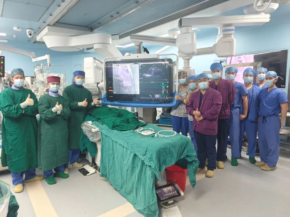 This is a Multidisciplinary Team at West China Hospital
Source: interviewee
Surgery Cannot Wait — But Which Approach?
Prof. Hu recalls vividly the first time he met Ms. Zhang. “Her condition was very poor, and surgery was urgent. But the patient and her family were adamant: only minimally invasive.”
If the heart is imagined as a pump, the mitral valve is the gate that ensures one-way flow. When it fails, the heart must work harder, and over time this leads to heart failure.
Ms. Zhang’s problem was exactly that. Seven years earlier, she had undergone surgery at West China Hospital, with the chest opened through a median sternotomy to receive a brand-new bioprosthetic mitral valve. Bioprosthetic valves, however, have an unavoidable shortcoming: natural aging.
Prof. Hu explained that, under years of blood flow and constant cardiac motion, bioprosthetic valves gradually calcify and thicken; in patients with metabolic conditions such as hyperlipidemia or diabetes, this process accelerates.
In the past year, Ms. Zhang’s bioprosthetic valve had deteriorated rapidly. She experienced worsening chest tightness and shortness of breath, becoming unable to walk more than a few steps without gasping. In the mornings, her eyelids were swollen; throughout the day, she was bloated; her legs were so swollen they glistened, leaving deep pits when pressed. Examination showed severe bioprosthetic valve stenosis, with a mean transvalvular gradient as high as 30 mmHg. Blood was pooling in the heart — unable to flow in or out — and cardiac functional reserve was nearly exhausted.
A second operation to replace the failed bioprosthetic valve was her only chance of survival.The surgical options included repeat open-chest surgery, thoracoscopic surgery, or an interventional approach. One path after another, however, was soon blocked.
At the mention of “open chest,” the whole family hesitated. The shadow of her previous surgery had long hung over them: Ms. Zhang had recovered slowly, enduring chest pain for years, and even now, in windy or rainy weather, she still felt a dull ache.
Moreover, a second open-chest operation carried far higher risk than the first — severe adhesions in the chest and pericardium were likely, and dissection could easily cause major bleeding, with blood loss of hundreds or even more than a thousand milliliters being common. Ms. Zhang said bluntly: “If I have to go under the knife again, then forget it.”
Thoracoscopic surgery was also unsuitable. On the one hand, adhesions from the previous open-chest surgery would also constrain thoracoscopic maneuvers; on the other hand, “thoracoscopic surgery requires cardiac arrest and cardiopulmonary bypass, which may worsen pulmonary edema and inflammation,” Prof. Hu explained. The patien’s lungs had been congested long-term, and she had already developed severe pulmonary hypertension with very poor pulmonary function; if this route were taken, she would likely require prolonged ventilator support after surgery, with a sharply elevated risk of infection.
Cases are not rare in clinical practice in which the heart operation succeeds but the patient cannot leave the table because of infection and pulmonary failure.
“My initial assessment was that there was a 70% possibility of an interventional approach, and we could choose the femoral vein–transseptal access for transcatheter mitral valve-in-valve (MVIV) replacement,” Prof. Hu said.
As workup deepened, however, a fatal “trap” emerged. Before the procedure, the team performed a millimeter-level three-dimensional reconstruction of the patient’s heart based on contrast-enhanced CT, simulating the post-implant configuration and repeatedly rehearsing the position and precision of the new valve leaflet against the old one.
Ms. Zhang’s reconstruction results were precisely the most dangerous category: with conventional transcatheter mitral valve replacement alone, left ventricular outflow tract (LVOT) obstruction would almost certainly occur.
“In conventional transcatheter valve replacement, the new valve is delivered into position without removing the old one. It’s like fitting a new door panel inside the existing door frame — the old door panel is naturally pushed aside. At the mitral position, however, if the original valve leaflet is pushed aside and blocks the channel below, it will obstruct the left ventricle’s outflow tract — the LVOT — causing LVOT obstruction,” Prof. Hu explained.
This kind of obstruction often strikes without warning. Once it occurs, blood cannot be pumped out normally, blood pressure drops sharply within seconds, ventricular fibrillation follows, and the patient can die instantly on the table. “There is almost no buffer. The surgeon doesn’t even have time to open the chest for rescue,” Prof. Hu said.
An operation that could not be delayed was caught in a “nothing works” deadlock. Where was the path forward?
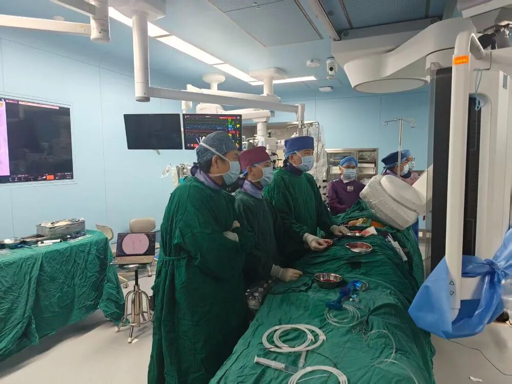 Prof. Hu’s Team During Surgery
Source: interviewee
“Threading a Needle” on a Beating Heart
After repeated rehearsal on the three-dimensionally reconstructed cardiac model, Prof. Hu’s team found a way forward: during MVIV, the BATMAN procedure — balloon-assisted translocation of the mitral anterior leaflet — was added. Specifically, the surgeon punctures the degenerated bioprosthetic valve with a wire and, like drawing a curtain, pulls part of the anterior leaflet toward the left ventricular wall. In this way, after the new valve is implanted, the old leaflet no longer obstructs the LVOT, effectively expanding the LVOT space and fundamentally eliminating the risk of obstruction.
This concept was introduced by a foreign team around 2020, but their approach used a transapical route, which required a small incision in the chest wall. Domestic attempts in China have since used both transapical and transaortic routes. For Ms. Zhang, however, a transapical approach would have required passing through cardiac muscle, causing partial myocardial fiber necrosis and scarring that could affect local contractile function; a transaortic approach, in contrast, required a long puncture distance, with a higher risk of damaging the aortic valve.
Ms. Zhang needed a less invasive and more reliable approach. Prof. Hu’s team ultimately decided to complete the entire procedure on a beating heart via the femoral vein–transseptal access.
How difficult is this? Imagine holding a very long wire and trying to pierce a thin sheet of paper fluttering in the wind, several meters away on an outer wall. Even more daunting, the surgical field is full of “minefields”; the slightest deviation could cause the wire to puncture a hole in the interventricular septum. Prof. Hu has indeed heard of such failed cases.
Asked why he dared to undertake this unprecedented-in-China operation, Prof. Hu said it was thanks to thorough preoperative planning and rehearsal, solid technical reserves, and seamless team coordination.
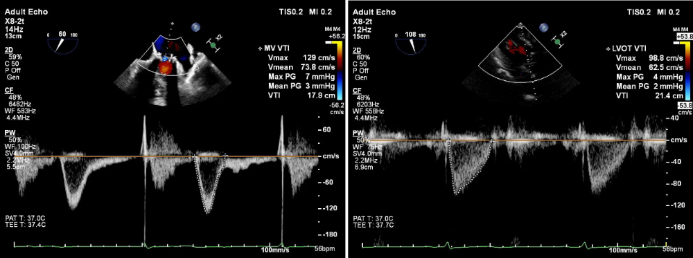 Left: Post-op Mitral Inflow Velocity and Gradient; Right: Post-op LVOT Velocity and Gradient, both Showing Marked Improvement
Source: interviewee
The anesthesiology team led by Prof. Song Haibo, chief physician of the Anesthesia and Surgery Center at West China Hospital, played a key role. Before surgery, the two teams repeatedly rehearsed on the cardiac model, refining details down to where the wire should pause, which views ultrasound should acquire, and how to evaluate the outflow tract before and after puncture.
“We agreed preoperatively that if the ultrasound could not provide clear imaging at a key location, we would not take the risk and would not gamble on luck,” Prof. Hu recalled. During the operation, the transesophageal echocardiography (TEE) guidance from Prof. Song’s team was, in his words, “perfect.” “Once the wire was inside, I held my breath, my hand trembling slightly with the patient’s heartbeat, and on Prof. Song’s command, I applied just enough force — and the wire went through to the exact spot.”
It was through this step-by-step discipline that the team completed, in order, transseptal puncture, leaflet puncture, balloon dilation, and transcatheter valve implantation. Immediate post-op echocardiography showed the new valve stably in place, opening and closing normally, with no sign of outflow tract obstruction. The key operative steps took about 2.5 hours, with total blood loss of about 10 ml. “That’s just the blood from the femoral artery and vein puncture at the groin — for cardiac surgery, it’s essentially negligible.” In Prof. Hu’s view, the minimal blood loss is, in itself, direct evidence of the team’s precision and coordination.
High-Difficulty Cardiac Surgery Is Becoming “Routine”
Ms. Zhang’s postoperative recovery was, in a word, dramatic.
Once the obstruction and perfusion issues were resolved, intraoperative parameters improved markedly, pulmonary hypertension eased substantially, and cardiac function improved almost immediately.
The anesthesia team assessed that she could be extubated in the operating room and transferred to a general ward directly. The next day, she was out of bed and walking on her own; by postoperative day 7, she was discharged. The swelling in her legs, which had been so severe, had visibly resolved, and she felt lighter overall. Happily, she said she would go square dancing at home.
“Generally, cardiac interventional patients can be discharged three to four days after the procedure. Ms. Zhang was in poorer overall condition, and her family was also concerned, so we kept her for two more days,” Prof. Hu said. Such a recovery speed, he added, is, in fact, “routine”in the department.
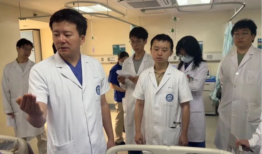 Prof. Hu’s Team on Ward Round
Source: interviewee
Behind this “routine” lies meticulous planning at every step by a large team.
Prof. Hu counted that, end to end, dozens of people were involved in Ms. Zhang’s operation: the Department of Cardiovascular Surgery led the procedure and operative planning, with three surgeons at the table; the nursing team handled pre- and post-op care, psychological support, and monitoring; Prof. Song’s team provided intraoperative ultrasound guidance; and the anesthesia team closely guarded the most dangerous steps of induction and intubation.
During surgery, the perfusionist was on standby throughout, ready to initiate emergent open chest if the interventional approach failed; the imaging team was responsible for preoperative data acquisition and cardiac model reconstruction; and the equipment department rapidly mobilized consumables.
“Complex and difficult cardiovascular surgery follows the ‘bucket principle’ — if even one plank is short, the patient may not leave the table,” Prof. Hu reflected.
He also observed that, in recent years, high-risk patients like Ms. Zhang, for whom traditional approaches offer no solution, are becoming increasingly common. This is closely related to the proliferation of heart-valve replacement and other cardiac procedures over recent decades; large numbers of patients are now reaching “bioprosthetic valve expiration” and need secondary intervention.
This is a new challenge for clinical practice, and it calls for more ingenious solutions. This first-in-China operation validates the safety and efficacy of transseptal MVIV combined with the BATMAN procedure in high-risk patients, and also demonstrates the value of domestic transcatheter valve devices in anatomically complex settings.
Prof. Hu is confident that, as clinical experience accumulates, such innovative procedures will benefit more patients with complex heart disease, especially the high-risk elderly with extremely unfavorable anatomy.

