


More than a century after modern neuroanatomy emerged, and 28 years after the internal nerve network of the penis was mapped, researchers in 2026 have finally produced the first detailed map of the human clitoral nerve network.
The scans show unprecedented detail. Five large tree-like nerve branches project from deep within the clitoral glans toward the surface, supplying a complex sensory network. For Dr. Ju Young Lee of Amsterdam UMC, who led the work, the striking point was not only what the scans revealed, but why such a seemingly basic anatomical task took so long.
"Female sexuality has been socially taboo, and that has obstructed scientific research."Dr. Lee said. The findings, released on bioRxiv in late March, suggest that some long-held medical assumptions about clitoral anatomy have been wrong.
A different picture inside the clitoral glans
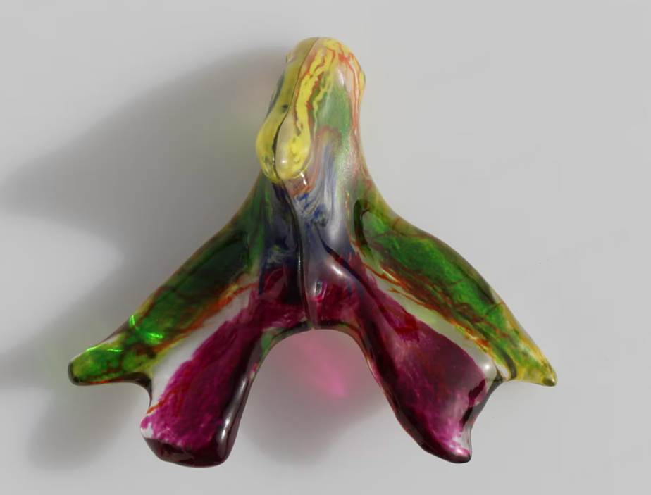
Dr. Lee's team found that the main sensory nerve, the dorsal nerve of the clitoris, branches from the pudendal nerve, travels along the internal erectile framework of the clitoris, and enters the clitoral body before extending into the glans.
Earlier anatomical studies had suggested that the nerve gradually thinned and disappeared after entering the glans. The new scans show the opposite: the nerves remain robust. Inside the glans, they divide into five large arborizing trunks and project densely from the center toward the surface. These trunks measured roughly 230 to 700 micrometers in diameter, with left and right branches supplying the corresponding half of the glans.
Dr. Georga Longhurst, head of anatomical sciences at St George's, University of London, said she was especially fascinated by the high-resolution images of the glans because such terminal branches cannot be seen in ordinary dissection.
The study used a circular particle accelerator nearly one kilometer in circumference to generate specialized X-rays. The team scanned the pelvic regions of two donated bodies and created micrometer-level 3D images, a level of detail beyond conventional dissection or MRI for the fine sensory fibers involved.
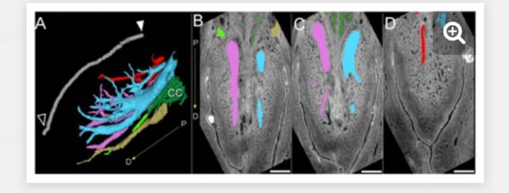
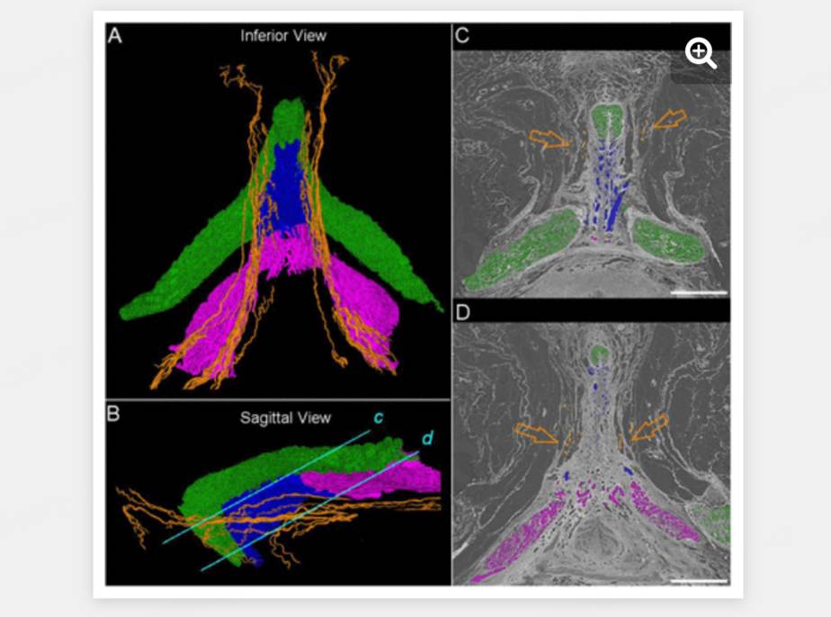
The scans also showed that some branches extend beyond the glans to tissue above the pubic bone, while others reach the clitoral hood. Another nerve, the posterior labial nerve, previously known to supply the labia majora and minora, also sends branches to both sides of the clitoral body. In other words, clitoral sensation appears to be covered by at least two intersecting neural systems rather than one isolated pathway.
Revisiting decades of anatomical bias
For decades, the nerve supply of the glans penis has been documented, while the clitoral glans remained poorly studied. Until a 2008 revision, Gray's Anatomy described the clitoris mainly as the homolog of the penis.
Although the penis and clitoris arise from the same embryonic structure, the genital tubercle, that framing treated the clitoris as a smaller version of a male organ. More recent work has shown that the clitoris is at least twice as large as older anatomy textbooks described, and its nerve density may be at least six times that of the penis.
The neglect reflects both social attitudes and clinical economics. In the mid-19th century, gender bias and emerging scientific theories diminished interest in female pleasure. Early 20th-century psychoanalytic ideas further marginalized clitoral orgasm. As obstetrics and gynecology became formalized, the field focused on pregnancy, delivery, and infertility, while sexual function was often dismissed as psychological.
By contrast, detailed nerve mapping of the penis was accelerated by the need to avoid erectile dysfunction after radical prostate cancer surgery. The large clinical demand created funding, prestige, and career incentives that clitoral research did not receive until pelvic floor repair, cosmetic gynecology, and gender-affirming surgery became more prominent.
Why the map matters in surgery
The immediate clinical significance lies in pelvic operations, including cancer treatment, reconstructive surgery, cosmetic procedures, and gender-affirming surgery. Without a complete understanding of clitoral anatomy, surgeons may inadvertently injure sensory nerves.
Pelvic organ prolapse repair, for example, takes place near the pudendal nerve, whose terminal branch is the dorsal nerve of the clitoris. In labiaplasty, surgeons often define a 'safe zone' based on earlier cadaveric dissection. Dr. Lee's team found that dorsal clitoral nerve branches extend into broader areas, meaning some procedures may be operating in regions previously assumed to be nerve-sparing.
The findings also matter for women who have undergone female genital mutilation. The research team noted that about 230 million women worldwide have been subjected to the practice, including removal of the visible clitoris and parts of the labia in 30 countries across Africa, the Middle East, and Asia. For surgeons performing reconstruction, knowing the sensory nerve pathways is essential.
The study remains an early step. It used only two pelvic samples from postmenopausal donors and did not examine broader age groups or autonomic nerves involved in sexual arousal. Dr. Lee said the work establishes an anatomical foundation, but future studies should expand the sample base and explore the functional and molecular characteristics of clitoral innervation.

