


Nearly 100 medical professionals spent 24 hours completing a face transplant. On February 2, 2026, Vall d'Hebron University Hospital in Barcelona, Spain, announced the news.
Reportedly, this was the world's first face transplant using a donor who had undergone assisted dying. Spain has reported six face transplants of varying extent to date, half of them performed at Vall d'Hebron University Hospital. Apart from the world's first full-face transplant in March 2010, this was the hospital's third such procedure.
The hospital stressed that the breakthrough in this case lay not only in the unusual donor source, but also in technical innovation. For the first time, the surgical team was able to perform complete preoperative 3D modeling for both donor and recipient, and used a near-infrared laser imaging device, continuous neurophysiological monitoring, and other technologies to improve postoperative recovery of facial function and sensation.
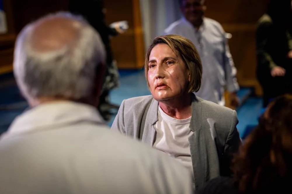
Only four months after surgery, the patient, a woman in her 50s, was already able to chew with her new face, speak, and make facial expressions with ease. Looking in the mirror, she found that the face was "growing more and more like my original self."
Face transplantation is the last treatment option
The story behind the operation began in an absurd and brutal way.
In July 2024, while on holiday, the patient was bitten by a small insect and subsequently developed a fatal "flesh-eating bacterial infection." The pathogen rapidly caused necrotizing fasciitis, leaving her in a coma for four months as she was treated in intensive care units at three different hospitals.
When she finally escaped death, the price was devastating destruction of half her face: she had lost half of her nose, and her lips, maxilla, part of her tongue, and facial muscles, including the orbicularis oculi that controls the eyelids, were almost completely destroyed. In addition, she lost her sense of smell, could not eat or breathe normally, and had extreme difficulty speaking.
"I saw many doctors, and all of them said this face could not be saved. Only Vall d'Hebron University Hospital gave me hope," the patient recalled.
After evaluating her, Dr. Joan-Pere Barret, head of the Plastic Surgery and Burns Department at Vall d'Hebron University Hospital, proposed the only treatment option: transplantation of the central facial region (type I facial defect).
"As human beings, we may suffer all kinds of physical disability, but facial disfigurement is among the most devastating. It not only deals a severe blow to the patient's mental health and social functioning, but also affects basic vital abilities such as speaking, eating, and breathing," the hospital said in a statement.
In general, face transplantation is used for patients who have extensive loss of facial tissue due to disease, burns, trauma, or congenital defects, and whose injuries cannot be repaired with conventional plastic surgery techniques.
According to Reuters and other media reports, as of February 2026, about 54 face transplants of varying extent had been completed worldwide, across about 20 medical centers in 11 countries. Only seven centers had completed at least three procedures, and Vall d'Hebron University Hospital was one of them.
Such operations are rare not only because they are technically extremely demanding, but also because donors are exceptionally scarce.
Matching requirements for face transplantation are extraordinarily strict: the donor and recipient must be of the same sex, have compatible blood types, similar facial shape, close skin tone, comparable age, and matching head size. That means recipients often wait for years, and may never find a suitable donor.
That was precisely what made this case unusual. In December 2024, the hospital began searching for a face transplant donor for the patient. Around the same time, a woman with an incurable genetic disease contacted the hospital to express her wish to undergo assisted dying - in Spain, assisted dying has been legal since 2021 - and she also volunteered to donate her major organs and tissues, including her face.
"For someone who has decided to end her life, the final wish is to save a stranger and give that person a chance to begin again," hospital staff said. This decision also fundamentally changed the preparation process for transplantation.
Previously, donors for face transplantation came from brain-dead patients, and medical teams had to complete organ retrieval and transplantation within an extremely limited time window. Donation after assisted dying, by contrast, allowed ample time for preparation, enabling the surgical team to carry out detailed imaging examinations of both donor and recipient, accurately measure facial compatibility, and devise a meticulous surgical plan.
The surgery set multiple world firsts
The patient's surgery was successfully completed in the autumn of 2025.
In medicine, face transplantation is known as the "Mount Everest" of surgery. It belongs to vascularized composite allotransplantation (VCA) and requires simultaneous transplantation of skin, fat, muscle, bone, nerves, and blood vessels.
Some tissues require suturing at diameters under 0.2 mm. Under the microscope, the surgical team had to precisely connect these different tissue types and ultimately create a new face that was both viable and functional. In terms of complexity and technical difficulty, the operation represented the outer limit of surgery.
The hospital spent months preparing. Nearly 100 professionals were involved at different stages, spanning more than 10 departments, including Plastic Surgery and Reconstructive Microsurgery, Anesthesiology, Immunology, Psychiatry, Rehabilitation, Intensive Care, Clinical Neurophysiology, Radiology, and the 3D Technology Center.
The core surgical team included 17 plastic surgeons, all experts in reconstructive microsurgery, 10 specialty residents, and about 25 nurses. The ample preoperative preparation time allowed the team to use the most advanced digital technologies available at the time.
The Radiology Department performed high-precision CT scans on both donor and recipient. After the imaging data were sent to the 3D Technology Application Center, the biomedical team quickly constructed precise three-dimensional digital models to help the surgical team plan the procedure.
"More than 10 years ago, when we performed the world's first full-face transplant, we had no 3D technology. More than 30 people relied entirely on experience and the naked eye to compare the anatomy of two individuals. As a result, a small number of motor and sensory nerves were stitched in the wrong positions. After the operation, when the patient wanted to blink, the corner of the mouth moved instead," the surgical team recalled.
With 3D models, engineers were able to customize precise bone-cutting guides for both donor and recipient. Surgeons only had to fit the guides onto the bone and cut along the preset slots to ensure exact cutting angle and depth, allowing donor and recipient bones to match with millimeter-level precision.
Using these models, the doctors could repeatedly rehearse the details of the procedure before surgery, almost like assembling building blocks, and fully prepare for any anatomical obstacle that might arise.
In addition, biomedical engineers used the models to customize a translucent silicone mask for the donor, to reconstruct the donor's appearance after surgery, "as the highest expression of respect for the donor and the donor's family."
The innovation in this face transplant was also reflected in the early adoption of high technology and real-time monitoring of surgical results. For example, this was the world's first face transplant in which a clinical neurophysiologist took part.
For the patient, normal anatomical structures had been severely destroyed and wrapped in scar tissue.
At that point, the surgeons were like electricians connecting wires, while the clinical neurophysiologist was like an inspector holding a multimeter. Through intraoperative electrical stimulation and electromyography (EMG) monitoring, the neurophysiologist could observe faint muscle contraction responses, helping the surgeons accurately locate specific small nerve branches within chaotic anatomy and avoid secondary damage caused by misconnection.
After donor and recipient nerves had been sutured, the neurophysiologist could also monitor electrical signal conduction in real time and make a preliminary intraoperative assessment of the potential for neural functional reconstruction, providing baseline data for postoperative rehabilitation.
According to the hospital, this was also the world's first time a near-infrared laser imaging device had been used during a face transplant to monitor facial perfusion.
After the microsurgical team completed the sutures, a fluorescent dye was injected intravenously. Under a special device, the dye became visible, allowing doctors to immediately see on a screen whether blood was effectively perfusing every terminal branch of the transplanted face. If insufficient perfusion was found, vascular anastomoses could be adjusted at once, instead of waiting until tissue necrosis appeared days after surgery.
"Our first face transplant patient spent more than a year in rehabilitation after surgery. The face was swollen and stiff for months, and at one point the patient could not even close the eyes. With the support of new technologies, however, this patient recovered facial touch, temperature perception, and other functions just over three months after surgery," the surgical team said.
Looking back at the three face transplants performed at the hospital, hospitalization time has steadily shortened: the first patient in 2010 remained hospitalized for three months after surgery, the second patient in 2015 for two months, and this patient went home after only one month.
Using photographs to awaken facial muscle memory
The great success of this face transplant also depended on comprehensive support from the multidisciplinary team at Vall d'Hebron University Hospital.
The anesthesiology and recovery team used advanced intraoperative monitoring and precise hemodynamic control to maintain circulatory stability throughout the 24-hour operation, control bleeding, perform metabolic resuscitation, and manage complex airway issues, laying the foundation for safe surgery.
In the early postoperative stage, before neural connections were fully established, the patient's face was in a hypotonic state and she could not make any expressions or movements. Physicians in Physical Medicine and Rehabilitation used mirrors, materials with different textures, and preoperative photographs to help her recall facial motor memory, rebuild facial visual perception, and stimulate recovery of neural function.
The rehabilitation team also developed a home facial rehabilitation plan for her and urged her to keep training at home to strengthen and activate more facial muscles. "I practice smiling at home. It feels a little strange, but I'm making progress every day," the patient said.
Meticulous psychological support was equally indispensable. Teams from Psychiatry, Clinical Psychology, and Social Work carried out comprehensive assessments and interviews with the patient and her family before and after surgery. Early postoperative psychological intervention was intended to help her process emotions, accept the change in appearance, and adapt to the effects of the surgery and immunosuppressive drugs.
Notably, about 25 of the nearly 100 professionals involved throughout the procedure were members of the nursing team. "Nurses were involved in every stage of the face transplant," the hospital said.
The hospital announced that the transplant had been highly successful, achieving both aesthetic results and functional recovery. "She can breathe and swallow smoothly, and she can once again eat with her mouth, something she could not do before the transplant."
According to the rehabilitation plan, the patient will enter the second phase of rehabilitation at six months after surgery, when psychological support will focus on helping her reenter society and live independently. But only four and a half months after surgery, she had already stepped outside, gone to her favorite cafe, and sat in the winter sun drinking coffee.
"I believe that by the first anniversary of the surgery, my life will be completely back to normal," the patient said.

