


“Standing Just 158 cm Tall, Yet Bearing a Giant 45 kg Tumor: A Chief Specialist from a Top-Tier Hospital Recounts the Diagnostic and Treatment Process”
A tumor weighing as much as 45 kg had grown inside a woman just 158 cm tall. Recently, Hunan Cancer Hospital admitted this extraordinary case.
Ouyang Qiang, Director of Gynecologic Oncology II at Hunan Cancer Hospital, told Medical界 that the patient, Ms. Wang, is 48 years old and has undergone multiple surgeries for aggressive angiomyxoma. This rare tumor typically affects women of childbearing age, is locally invasive, and has a high rate of recurrence.
Sixteen years ago, Ms. Wang had surgery for this disease at Hunan Cancer Hospital, but she did not return for regular follow-ups. Eight years ago it recurred and gradually enlarged. Before coming back to Hunan Cancer Hospital this time, she had sought care at multiple hospitals across China, but was turned away everywhere due to the high surgical difficulty and risks involved.
“Because the tumor occupied most of her abdominal and pelvic cavities, she suffered shortness of breath and even cardiac and pulmonary impairment,” Ouyang explained. After multidisciplinary consultations, preoperative preparation, intraoperative management, and postoperative care, the team successfully removed the tumor.
Tumor Over 50 cm, Weighing 45 kg
Ouyang still remembers his first glimpse of Ms. Wang. “When we opened her clothing, her abdomen was distended and shiny from tumor compression—truly a shocking sight.” At the time, her overall condition was poor: at 158 cm tall and roughly 170 jin (including the tumor), her body mass index was far below normal, indicating severe malnutrition. Her hemoglobin was just 46 g/L (normal: 110–150 g/L), reflecting profound anemia.
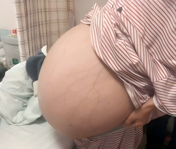
Data and images sourced from Hunan Cancer Hospital.
Ms. Wang had already undergone three prior surgeries for aggressive angiomyxoma. Her surgical history began in 2002, when during a cesarean section at a county hospital in Huarong, Yueyang, a perineal mass was discovered and removed but not sent for pathology. Between 2003 and 2005 the tumor recurred in her pelvis; a local hospital excised the mass, and pathology confirmed a mesenchymal tumor.
In 2009 she returned to Hunan Cancer Hospital with masses in the vulva and pelvis—an approximately 10 cm intra-abdominal tumor that had severely adhered to her bladder, blood vessels, pelvic wall, and ureter—undergoing another excision.
Around 2017 she began to experience abdominal distension and pain; imaging then showed the tumor had grown to 20–30 cm. Doctors warned that surgery carried high risks, and financial hardship prevented timely treatment.
Over the following years, she and her family traveled from hospital to hospital, but most declined her case due to its complexity. She endured at home until this year, when the mass became so large that she had difficulty breathing, eating, and even lying down, prompting her to seek help again at Hunan Cancer Hospital.
Disease Characteristics and Surgical Challenges
Aggressive angiomyxoma (AAM) is a rare benign mesenchymal tumor that arises in the pelvic and perineal regions of women of reproductive age, characterized by local infiltration and a high recurrence rate. Though benign and with low metastatic potential, its continuous growth can severely compromise quality of life, causing symptoms such as respiratory distress.
At presentation, Ms. Wang’s anemia and malnutrition placed her at risk for organ failure if the tumor continued to grow. Compared to more malignant gynecologic tumors, AAM has a relatively favorable prognosis, but its slow, painless enlargement often means that by the time of diagnosis, tumors are quite large.
Their infiltrative nature makes complete surgical excision difficult.Ouyang outlined three main preoperative concerns:
Poor patient condition: Malnutrition and anemia reduce immunity and strain cardiac function.
Extensive tumor burden and dense adhesions: Prior surgeries had caused severe scarring, and the tumor was tightly adherent to surrounding organs, raising the risk of intraoperative injury.
High vascularity and size: A richly blood-supplied, large tumor poses a major hemorrhage risk, and repairing the abdominal wall afterward to maintain organ position and function is challenging.
Multidisciplinary Preparation and Surgical Strategy
The hospital convened four full-hospital multidisciplinary meetings involving dozens of departments: interventional radiology, hepatobiliary-colorectal surgery, urology, breast and reconstructive surgery, radiology, anesthesia, critical care, nutrition, and more. Key measures included:
Interventional embolization prior to surgery to reduce intraoperative bleeding
Angiography and 3D-printed models to map tumor relations to adjacent structures
Nutritional support with supplements and feeding guidance
Anesthetic hemodynamic planning to handle potential blood loss
Reconstructive planning by plastic surgeons to restore abdominal wall integrity
Historically, when the tumor was only 10 cm, surgery resulted in 1,600 mL of blood loss; this time, the team estimated up to 10,000 mL. Thanks to embolization, however, intraoperative hemorrhage was well controlled.Preoperative imaging also revealed cystic areas within the tumor, making percutaneous drainage a viable tactic to decompress the abdomen and mitigate decompression syndrome during resection.
By draining the cystic fluid, the solid component was reduced to 38 jin, easing the operative burden—yet dense adhesions to bowel, bladder, vessels, and ureter remained.
Surgical Procedure and Postoperative Care
Under the leadership of Professor Mao Xianhai, Vice President and Head of Hepatobiliary-Colorectal Surgery, the team employed advanced instruments to meticulously dissect and remove the mass.
Urologists then placed ureteral stents to protect the urinary tract, while colorectal surgeons separated and preserved the intestines. Anesthesia and interventional teams worked in concert to manage decompression effects, and despite an estimated 1,500 mL of blood loss, Ms. Wang was stable and transferred to the ICU for three days of observation due to low blood pressure.
Given AAM’s high recurrence risk, the team obtained intraoperative samples to culture tumor organoids and devised an adjuvant therapy plan. Their goal is to combine surgical and research efforts to customize further treatment if recurrence occurs, thereby extending survival and improving quality of life.
Ms. Wang has now been discharged and continues postoperative care at a local hospital in Yueyang. Encouragingly, her anemia and nutritional status have markedly improved, and she has gained weight.
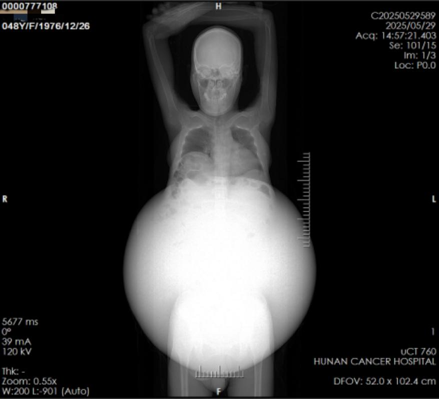 Preoperative imaging data of the patient
Institutional Expertise and Experience
Hunan Cancer Hospital is one of China’s first tertiary-level cancer specialty hospitals and a designated national cancer regional medical center. Its Gynecologic Oncology Department II, founded in 2005, is among the hospital’s earliest subspecialty units, and has long been a national leader in the diagnosis, treatment, and research of gynecologic tumors. While the department has treated other AAM cases, none approached the size of Ms. Wang’s tumor.
Reflecting on this rare and challenging case, Ouyang noted that although he had concerns before surgery, the patient’s trust in the specialists imposed a duty to do everything possible. “Our hospital’s ‘three minimal invasions and three preservations’ philosophy—minimally invasive, endoscopic, interventional; preserving efficacy, organs, and patient comfort—guided the entire process, and has given us invaluable experience in managing such complex conditions.”

