


Introduction
Non-obstructive hypertrophic cardiomyopathy (nHCM) does not produce a significant left ventricular outflow tract (LVOT) gradient, but myocardial hypertrophy, diastolic dysfunction, atrial remodeling, and myocardial ischemia still cause exertional dyspnea, fatigue, and reduced exercise tolerance, while patients face long-term risks of heart failure progression, atrial fibrillation, and sudden cardiac death. Compared with obstructive HCM (oHCM), current treatments for nHCM (beta-blockers, calcium channel blockers) only relieve symptoms, and targeted drugs that meaningfully improve cardiac structure and function have long been lacking.
The European Society of Cardiology (ESC) Congress 2026 was held in Munich, Germany, from August 28 to 31, 2026. ACACIA-HCM is the first phase 3 trial to achieve both primary endpoints in symptomatic nHCM, with aficamten improving both peak oxygen consumption (pVO2) and the Kansas City Cardiomyopathy Questionnaire Clinical Summary Score (KCCQ-CSS) versus placebo at week 36, making it a major topic in the cardiomyopathy field at ESC 2026. During the Late-Breaking session at ESC 2026, Prof. Sheila Hegde of the University of Texas Southwestern Medical Center delivered a dedicated interpretation of the pre-specified exploratory echocardiographic subgroup analysis of ACACIA-HCM, with the related results published in Circulation that same day. The subgroup analysis systematically evaluated echocardiographic data from all 517 participants to characterize the benefit of aficamten on cardiac structure and function in patients with nHCM.
This article summarizes the subgroup analysis’s core findings on cardiac structure, cardiac function, safety, and correlation with the primary clinical endpoints, and discusses its clinical application and value.
Article Key Points
- ACACIA-HCM is a multicenter, randomized, double-blind, placebo-controlled phase 3 trial in 517 adults with symptomatic nHCM randomized 1:1 to aficamten or placebo, with a 36-week treatment period extendable to 72 weeks.
- At week 36, aficamten reduced maximal wall thickness (MWT) by 0.13 cm (95% CI -0.20 to -0.07; p<0.001) and septal wall thickness by 0.12 cm, with reversal extending to -0.20 cm at end of treatment; reduction in inferolateral wall thickness did not reach statistical significance.
- LAVI improved by 1.3 mL/m² at week 36 and by 1.7 mL/m² at end of treatment (p=0.022); each 50% reduction in NT-proBNP was associated with a 1.06 mL/m² LAVI decrease (p=0.035), consistent with coordinated improvements across cardiac load, structure, and biomarkers.
- Diastolic function improved significantly, with septal e’ increasing by 0.7 cm/s and lateral e’by 0.9 cm/s (both p<0.001); the proportion with normal septal e’ rose from 26% to 39% and normal lateral e’ from approximately 52% to 64% at week 36.
- LVEF declined by 4.6% at week 36 (95% CI -5.5 to -3.6; p<0.001) but remained >50% in the vast majority of participants; 10.5% had reversible LVEF <50% and 2.7% required dose suspension, with rapid recovery after 4-week washout.
Pre-specified Exploratory Analysis: Echocardiographic Data From All 517 Participants Over 72 Weeks
ACACIA-HCM is a multicenter, randomized, double-blind, placebo-controlled phase 3 trial in which 517 adults with symptomatic nHCM were randomized 1:1, with a 36-week treatment period extendable to 72 weeks. The subgroup analysis drew on echocardiographic data from all 517 participants through week 36 and matched the 72-week extended-treatment results.
Baseline characteristics: mean age 55 ± 16 years; female 54%; White 75%, Asian 13%; paroxysmal atrial fibrillation 28%; background beta-blocker use 77%; New York Heart Association (NYHA) class II/III 68%/31%; mean pVO2 18.0 ± 5.2 mL·kg⁻¹·min⁻¹; cavity obstruction approximately 9%. Echocardiographic baseline showed mean left ventricular ejection fraction (LVEF) 68 ± 4%, maximal wall thickness 2.3 ± 0.5 cm, left atrial volume index (LAVI) 36.8 ± 10.9 mL/m², and typical diastolic abnormalities (reduced septal e’, elevated septal E/e’).
Cardiac Structural Changes
Significant Reversal of Myocardial Hypertrophy: Reductions in Maximal Wall Thickness and Septal Thickness
Based on these baseline features, the exploratory analysis evaluated the treatment effect of aficamten across two dimensions: cardiac structure and cardiac function. As myocardial hypertrophy is the core pathologic change in nHCM, at week 36 aficamten reduced maximal wall thickness (MWT) versus placebo by 0.13 cm (95% CI -0.20 to -0.07; p<0.001), and by 0.20 cm at end of treatment (EoT) (95% CI -0.30 to -0.10). Septal thickness decreased by 0.12 cm at week 36 and by 0.20 cm at EoT (both p<0.001). Reduction in inferolateral wall thickness did not reach statistical significance.
Table 1. ACACIA-HCM Subgroup Analysis: Treatment Differences of Aficamten on Myocardial Hypertrophy and Left Ventricular Volume
|
Parameter |
Baseline (Afarcastat vs Placebo) |
Treatment Difference at Week 36 |
P Value |
EoT Treatment Difference |
|
Maximal Wall Thickness (cm) |
2.2±0.5 vs 2.3±0.6 |
-0.13 (-0.20, -0.07) |
<0.001 |
-0.20 (-0.3, -0.1) |
|
Interventricular Septal Thickness (cm) |
1.6±0.4 vs 1.6±0.4 |
-0.12 (-0.17, -0.08) |
<0.001 |
-0.20 (-0.2, -0.1) |
|
Inferolateral Wall Thickness (cm) |
1.0±0.3 vs 1.0±0.3 |
-0.03 (-0.07, 0.01) |
0.190 |
-0.02 (-0.06, 0.02) |
|
Left Ventricular End-Diastolic Volume Index (LVEDVi) (mL/m²) |
35.4±7.2 vs 34.6±7.0 |
+4.1 (2.8, 5.4) |
<0.001 |
+3.2 (1.9, 4.5) |
|
Left Ventricular End-Systolic Volume Index (LVESVi) (mL/m²) |
11.4±3.2 vs 11.0±2.7 |
+3.2 (2.5, 4.0) |
<0.001 |
+2.8 (2.1, 3.6) |
|
LVEF (%) |
67.9±4.0 vs 68.4±3.4 |
-4.6 (-5.5, -3.6) |
<0.001 |
-4.0 (-4.9, -3.1) |
|
Parameter |
Baseline (Afarcastat vs Placebo) |
Treatment Difference at Week 36 |
P Value |
EoT Treatment Difference |
The magnitude of this reverse remodeling is similar to that observed with other cardiac myosin inhibitors (CMIs) in studies such as ODYSSEY-HCM, suggesting that aficamten may also reverse myocardial hypertrophy; future cardiac magnetic resonance (CMR) substudies are expected to provide independent confirmation of these structural changes.
Stabilization of Left Atrial Remodeling: LAVI Improvement Linked With NT-proBNP Reduction
Alongside reversal of myocardial hypertrophy, the effect of aficamten on atrial remodeling is also noteworthy. LAVI is a key indicator of atrial remodeling and chronic elevation of left ventricular filling pressure, and was a pre-specified secondary endpoint in ACACIA-HCM. At week 36, LAVI in the aficamten group was reduced by 1.3 mL/m² versus placebo (95% CI -2.6 to 0.04; p=0.06); extended treatment to EoT (median 49 weeks) widened the difference to 1.7 mL/m² (p=0.022).
LAVI improvement was linked with N-terminal pro-B-type natriuretic peptide (NT-proBNP) reductions: each 50% reduction in NT-proBNP was associated with a 1.06 mL/m² decrease in LAVI (p=0.035), a 1.01 decrease in septal E/e’ (p<0.001), and a 0.07 cm reduction in MWT (p=0.001). This coordinated improvement in cardiac load, structure, and biomarker may represent the imaging foundation for aficamten’s dual improvement in symptoms and cardiac function.
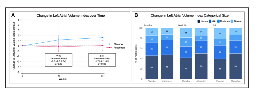 Fig. 1. Effect of Aficamten on Left Atrial Volume Index (LAVI) Over Time
Cardiac Function Improvement
Significant Improvement in Diastolic Function: Increased e’ Velocity and Reduced E/e’
Parallel to atrial remodeling, diastolic dysfunction is the earliest and most central pathologic feature of nHCM. Baseline data showed lateral e’ at the lower limit of normal, reduced septal e’ (5.1 ± 1.7 cm/s), and elevated septal E/e’ (15.4 ± 7.1), consistent with the spectrum of diastolic dysfunction defined in the 2025 American Society of Echocardiography (ASE) guidelines.
At week 36, aficamten increased lateral e’ by +0.9 cm/s versus placebo (95% CI 0.5 to 1.2) and septal e’ by +0.7 cm/s (95% CI 0.4 to 0.9), both p<0.001, with effects persisting at EoT. Peak E-wave velocity increased by 5.1 cm/s (p=0.001), consistent with the physiologic direction of improved myocardial relaxation and enhanced early filling.
Septal E/e’ declined significantly only at EoT (-1.4, p<0.001); lateral E/e’ did not reach significance at either timepoint, consistent with the more pronounced baseline abnormality on the septal side. Stratified by ASE criteria, the proportion with normal septal e’ rose from 26% to 39% at week 36, and normal lateral e’ from approximately 52% to 64%; mean E/e’ showed no between-group difference at week 36 because baseline values were largely normal, and although baseline abnormality rates for septal and lateral E/e’ were high, the between-group difference at week 36 also did not reach statistical significance. In summary, aficamten significantly improves mitral annular early diastolic velocity e’, and with extended treatment, septal E/e’ also declines.
Table 2. ACACIA-HCM Subgroup Analysis: Treatment Differences of Aficamten on Left Ventricular Diastolic Function Parameters
|
Diastolic Function Parameter |
Treatment Difference at Week 36 |
P Value |
EoT Treatment Difference |
|
Lateral e’Velocity (cm/s) |
+0.9 (0.5, 1.2) |
<0.001 |
+0.9 (0.5, 1.2) |
|
Septal e’Velocity (cm/s) |
+0.7 (0.4, 0.9) |
<0.001 |
+0.9 (0.6, 1.1) |
|
Peak E Wave Velocity (cm/s) |
+5.1 (2.2, 8.0) |
0.001 |
+2.9 (0.2, 5.5) |
|
E/A Ratio |
+0.13 (0.03, 0.23) |
0.009 |
+0.1 (-0.02, 0.2) |
|
Lateral E/e’ |
-0.3 (-0.9, 0.3) |
0.38 |
-0.5 (-1.1, 0.1) |
|
Septal E/e’ |
-0.7 (-1.5, 0.1) |
0.07 |
-1.4 (-2.1, -0.6) |
|
LAVI (mL/m²)
|
-1.3 (-2.6, 0.04) |
0.06 |
-1.7 (-3.2, -0.3) |
LAVI analyses excluded participants with atrial fibrillation or atrial flutter on baseline ECG.
Mild Decline in LVEF but Overall Preserved: Dose-Dependent and Reversible After Washout
In contrast to the improvement in diastolic function, aficamten, as a CMI, produced the expected mild decline in left ventricular systolic function. Aficamten dosing was titrated based on LVEF: starting at 5 mg with stepwise up-titration at weeks 2, 4, and 6 for participants with LVEF ≥60% (up to a maximum of 20 mg); LVEF 50-60% maintained dose, <50% dose reduction, <40% dose interruption. By week 8, more than 80% of participants were maintained on 15-20 mg.
With longer titration time, LVEF declined by 0.8% at week 2 (p=0.015) and by 3.4% at week 8 (p<0.001); the treatment difference stabilized at -4.6% (95% CI -5.5 to -3.6; p<0.001) at week 36 and was -4.0% at EoT. LVEDVi increased by 4.1 mL/m² at week 36 and LVESVi by 3.2 mL/m². Although LVEF fell to 60.9 ± 6.7%, the vast majority of participants retained LVEF in the normal range (>50%), with only 10.5% (27 cases) experiencing transient reversible LVEF <50% (of whom 2.7% required dose interruption); values returned rapidly to baseline after 4 weeks of washout (EoS -0.5%; p=0.21), suggesting the changes are reversible and controllable.
Imaging-Driven Clinical Benefit: Correlation With Clinical Endpoints
Do these structural and functional changes actually drive the observed clinical benefit? The analysis pre-specified regression analyses of clinical endpoints against changes in imaging metrics. In the aficamten group (the placebo group showed only isolated right ventricular metrics correlated with endpoint changes), multiple structural and diastolic improvements correlated significantly with the clinical endpoints, forming two pathways: first, improvement in septal e’ velocity correlated with both primary endpoints (pVO2, KCCQ-CSS) and the secondary endpoint NT-proBNP, suggesting that diastolic improvement is a core driver of dual clinical benefit; second, reduction in MWT correlated significantly with reductions in NT-proBNP and improvement in KCCQ-CSS, suggesting that hypertrophy reversal also underlies the benefit.
Table 3. Correlation Between Changes in Clinical Endpoints and Imaging Changes in Patients Receiving Aficamten
|
Clinical Endpoint Change |
Imaging Parameter Change |
β Coefficient (95% CI) |
P Value |
|
pVO₂ per 1-unit increase |
Septal e’ Velocity |
+0.08 cm/s (0.01, 0.14) |
0.022 |
|
KCCQ-CSS per 5-point increase |
Septal E/e’ |
-0.22 (-0.42, -0.03) |
0.025 |
|
KCCQ-CSS per 5-point increase |
Maximal Wall Thickness |
-0.02 cm (-0.03, -0.01) |
0.009 |
|
NT-proBNP Decrease by 50% |
Septal e’ Velocity |
+0.35 cm/s (0.20, 0.50) |
<0.001 |
|
NT-proBNP Decrease by 50% |
Lateral ‘ Velocity |
+0.25 cm/s (0.01, 0.48) |
0.043 |
|
NT-proBNP Decrease by 50% |
Septal E/e’ |
-1.01 (-1.57, -0.45) |
<0.001 |
|
NT-proBNP Decrease by 50% |
Maximal Wall Thickness |
-0.07 cm (-0.12, -0.03) |
0.001 |
|
NT-proBNP Decrease by 50% |
LAVI |
-1.06 mL/m² (-2.04, -0.07) |
0.035 |
In addition, in the aficamten group, changes in LVEF, left ventricular global longitudinal strain (GLS), and cardiac output did not correlate significantly with changes in the clinical endpoints. Stratifying by baseline atrial fibrillation status (LAVI analyses excluded participants with baseline atrial fibrillation), treatment effects for the remaining diastolic metrics showed no significant difference between atrial fibrillation subgroups, suggesting consistent benefit across rhythm subgroups.
Safety Profile: Reversible LVEF Decline and Mild Right Ventricular Effects
Beyond efficacy, the safety and reversibility of these cardiac function changes are equally important. LVEF decline is an expected pharmacologic effect of CMIs. Core laboratory and site evaluations showed consistent results (week 36 treatment differences of -4.6% and -4.0%, respectively, with no significant treatment-by-assessment interaction; p=0.29), confirming that the decline is robust and reproducible; absolute LVEF remained >50% in the vast majority during titration, with rapid recovery during washout (-0.5%; p=0.21), consistent with reversibility seen in prior CMI studies.
For the right ventricle, aficamten increased right ventricular end-diastolic area by 1.0 cm² and end-systolic area by 0.7 cm²; TAPSE decreased by -0.10 cm (p=0.005), RV s’ by -1.1 cm/s (p<0.001), and peak tricuspid regurgitation velocity by -10.4 cm/s (p=0.03), but absolute values at EoT remained within the normal range. The mild increase in right ventricular area was accompanied by mild decreases in systolic metrics and reduced tricuspid regurgitation velocity; the latter suggests a reduction in right ventricular systolic pressure (reflecting left atrial pressure); the mild synchronous decline in biventricular systolic function is consistent with the targeted pharmacologic action of CMIs.
From Symptom Control to Structural Reversal: The Clinical Value of Aficamten
Synthesizing these findings, the clinical value of aficamten in nHCM can be understood at three levels: pathologic mechanism, regulatory pathway, and clinical practice.
At the mechanism level, the ACACIA-HCM echocardiographic exploratory analysis is the first to confirm at the imaging level that aficamten may reverse myocardial hypertrophy, stabilize left atrial volume, and improve diastolic function, advancing nHCM management from “symptom control” toward a comprehensive approach of “structural reversal + functional improvement.” As a next-generation CMI, aficamten improves diastolic function through reducing the number of myosin heads entering the force-generating state, promoting cross-bridge detachment, and lowering myocardial energy expenditure; evidence that this mechanism operates in the absence of LVOT obstruction is the core contribution of this exploratory analysis.
At the regulatory level, building on aficamten’s existing approval for oHCM, the structural and functional improvement data from this exploratory analysis may serve as key imaging evidence for an nHCM indication submission, forming a complete evidence chain together with the dual primary endpoints and supporting updates to HCM guidelines, ushering the full HCM spectrum (obstructive and non-obstructive) into the molecularly targeted therapy era.
At the clinical practice level, improvement in septal e’ velocity correlated with all three clinical endpoints (pVO2, KCCQ-CSS, NT-proBNP) and may serve as the most sensitive early signal of treatment response; patients with more pronounced baseline septal E/e’ abnormalities and significantly elevated NT-proBNP appear to derive greater benefit.
Summary
The ACACIA-HCM echocardiographic exploratory analysis is the first pre-specified exploratory study to systematically evaluate CMI-related echocardiographic effects in symptomatic nHCM. For the first time, it provides a possible mechanism-based imaging interpretation of the clinical benefit of aficamten in the absence of LVOT obstruction, partially answering the long-standing question of how CMIs deliver benefit without LVOT obstruction, and upgrading the long-term management goal for nHCM to structural reversal plus symptom improvement plus functional enhancement.
References
[1] Ommen SR, Ho CY, Asif IM, et al. 2024 AHA/ACC/AMSSM/HRS/PACES/SCMR Guideline for the Management of Hypertrophic Cardiomyopathy. Circulation. 2024;149(23):e1239-e1311. doi:10.1161/CIR.0000000000001250.
[2] Arbelo E, Protonotarios A, Gimeno JR, et al. 2023 ESC Guidelines for the management of cardiomyopathies. Eur Heart J. 2023;44(37):3503-3626.
[3] Pelliccia F, Pasceri V, Limongelli G, et al. Long-term outcome of nonobstructive versus obstructive hypertrophic cardiomyopathy: a systematic review and meta-analysis. Int J Cardiol. 2017;243:379-384. doi:10.1016/j.ijcard.2017.06.071.
[4] Lu DY, Pozios I, Haileselassie B, et al. Clinical outcomes in patients with nonobstructive, labile, and obstructive hypertrophic cardiomyopathy. J Am Heart Assoc. 2018;7(5):e006657. doi:10.1161/JAHA.117.006657.
[5] Masri A, Coats CJ, Maron MS, et al. Aficamten for Symptomatic Nonobstructive Hypertrophic Cardiomyopathy: Primary Results of the ACACIA-HCM Trial. Presented at: ESC Congress 2026; August 28, 2026; Munich, Germany.
[6] Masri A, Maron MS, Bhatia A, et al. Aficamten for Symptomatic Nonobstructive Hypertrophic Cardiomyopathy. N Engl J Med. 2026. doi:10.1056/NEJMoa2603021.
[7] Hegde SM, Wang X, Bhatia A, et al. Effect of Aficamten on Cardiac Structure and Function in Patients with Symptomatic Nonobstructive Hypertrophic Cardiomyopathy. Circulation. 2026;154:00-00. doi:10.1161/CIRCULATIONAHA.126.082343.
[8] Nagueh SF, Sanborn DY, Oh JK, et al. Recommendations for the Evaluation of Left Ventricular Diastolic Function by Echocardiography and for Heart Failure with Preserved Ejection Fraction Diagnosis: An Update from the American Society of Echocardiography. J Am Soc Echocardiogr. 2025;38:537-569. doi:10.1016/j.echo.2025.03.011.
[9] Maron MS, Masri A, Nassif ME, et al; SEQUOIA-HCM Investigators. Aficamten for Symptomatic Obstructive Hypertrophic Cardiomyopathy. N Engl J Med. 2024;390:1849-1861. doi:10.1056/NEJMoa2401424.

