


Consider a scenario that invites assumption. A 41-year-old man with type 2 diabetes for 2 years and hypertension for 6 months had a health check in June 2024 showing a serum creatinine of only 82 μmol/L and an estimated glomerular filtration rate (eGFR) of 102.64 mL/min/1.73 m², with a completely normal urinalysis. Seven months later he presented after 1 day of dizziness. Serum creatinine had risen to 258 μmol/L, and on admission to 299 μmol/L, with an eGFR of 21.33 mL/min/1.73 m².
What had happened? A case report published in Medicine describes a presentation in which routine laboratory testing offered no answer until kidney biopsy revealed that the cause was not progression of diabetic nephropathy but a form of acute injury that is easily overlooked.
Article Key Points
• Serum creatinine rose from 82 to 299 μmol/L in 7 months in a 41-year-old man with type 2 diabetes
• Urinalysis was normal and no cause was visible; renal biopsy showed acute interstitial nephritis
• The only plausible trigger was an unidentified Chinese herbal decoction taken for 15 days
• Glomerular changes matched class IIa diabetic nephropathy in the 2010 Renal Pathology Society classification
• Prednisone 30 mg/d was started on January 15, 2025 and tapered to complete withdrawal by June 17
• Creatinine had already fallen to 272 μmol/L before steroids, so recovery was not attributable to them alone
• Glucocorticoids caused hyperglycaemia and insomnia, requiring premixed human insulin 30R
• At the last follow-up creatinine was 121 μmol/L and eGFR 63.23 mL/min/1.73 m²
• Unexplained acute kidney injury in diabetes warrants biopsy and a search for superimposed interstitial nephritis
 Fig. 1. Title Page of the Source Case Report in Medicine (Baltimore)
Clues in a Bowl of Unidentified Chinese Herbal Medicine
Detailed history taking brought a key detail to light. Over the 2 years of diabetes, the patient had taken metformin 0.5 g once daily without regular blood glucose monitoring, and for hypertension amlodipine 5 mg once daily, although he in fact took it on only about 4 days a week. One month before onset, dissatisfied with his sexual function, he was given a decoction of Chinese herbal medicine at a local traditional Chinese medicine clinic and took it twice daily for 15 consecutive days. In the preceding 3 months he denied any use of antibiotics, non-steroidal anti-inflammatory drugs, proton pump inhibitors, health supplements or contrast agents, and he had no fever, rash, arthralgia or urinary tract infection.
On admission his blood pressure was 163/117 mmHg with mild pitting oedema of both lower limbs. Laboratory tests on January 5, 2025 showed a serum creatinine of 299 μmol/L, an eGFR of 21.33 mL/min/1.73 m², a blood urea nitrogen (BUN) of 14.88 mmol/L, a urine albumin-to-creatinine ratio (UACR) of 56.58 mg/g and a glycosylated haemoglobin (HbA1c) of 7.7%. White blood cells were 10.22×10⁹/L, with eosinophils at 0.53×10⁹/L, accounting for 5.2%. Urinalysis was negative for protein and occult blood, with no white cell casts, red cell casts or eosinophils. Markers of infection, hepatitis viruses, autoantibodies (antinuclear antibody, anti-double-stranded DNA antibody and antineutrophil cytoplasmic antibody) and complement were all normal. Renal ultrasound showed mild bilateral renal enlargement, increased cortical echogenicity and loss of the corticomedullary differentiation, with no obstruction and no stones.
Neither the available laboratory results nor the ultrasound findings could define the cause, or distinguish it from the most obvious alternative, progression of diabetic nephropathy.
One Biopsy, Two Processes at Once
On January 10, 2025, kidney biopsy provided the answer. On light microscopy, 1 of 10 glomeruli was globally sclerosed; the remaining glomeruli showed mild diffuse mesangial cell and matrix proliferation with mild thickening of the glomerular basement membrane and no nodular sclerosis (Fig. 2). Tubular epithelial cells showed granular and vacuolar degeneration with loss of the brush border, with acute injury in approximately 70% of tubules and mild tubulitis in fewer than 5%. The interstitium was oedematous, with inflammatory cell infiltration in approximately 10% of the cortical area, predominantly lymphocytes with scattered plasma cells and a few eosinophils, and interstitial fibrosis with tubular atrophy of approximately 10%.
Immunofluorescence was negative for IgG, IgA and C3, with only segmental weak IgM positivity in the mesangium and albumin reabsorption droplets within tubular epithelial cells (Fig. 3). Electron microscopy showed homogeneous thickening of the glomerular basement membrane, with a mean of approximately 500 nm, foot process effacement in less than 25%, and no electron-dense deposits (Fig. 4).
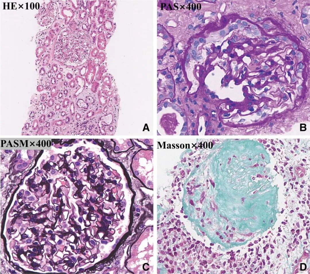 Fig. 2. Kidney Biopsy by Light Microscopy: Interstitial Oedema and Focal Inflammatory Cell Infiltration, with Mild Mesangial Proliferation and Thickening of the Glomerular Basement Membrane
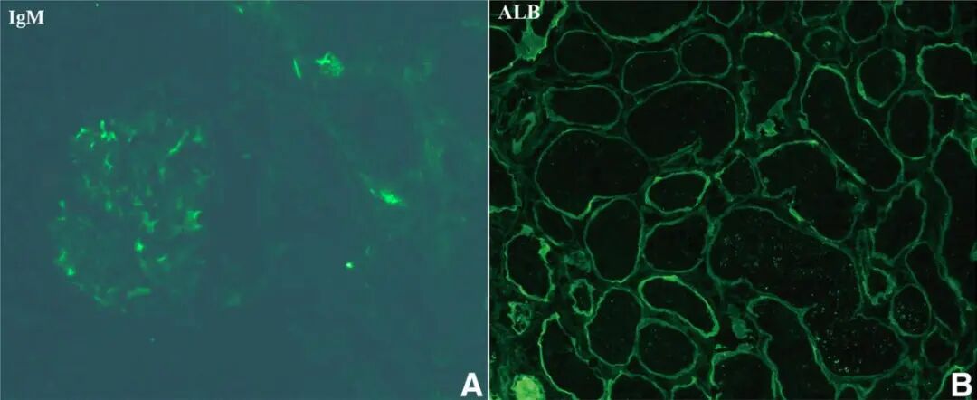 Fig. 3. Immunofluorescence: Segmental Weak IgM Positivity in the Mesangium, with Albumin Reabsorption Droplets in Tubular Epithelium
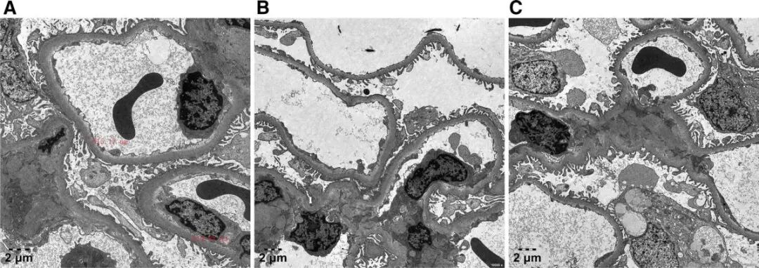 Fig. 4. Electron Microscopy: Homogeneous Thickening of the Glomerular Basement Membrane, Foot Process Effacement Not Exceeding 25%, and No Electron-Dense Deposits
Taken together, the combination of interstitial inflammatory cell infiltration and tubular injury is the pathological signature of acute interstitial nephritis (AIN). Negative immunofluorescence and the absence of electron-dense deposits on electron microscopy further excluded immune complex glomerulonephritis, and AIN was confirmed. The unidentified herbal decoction is the only plausible suspect, but its composition cannot be confirmed and can only be described as possibly related. The glomerular changes were consistent with class IIa diabetic nephropathy in the Renal Pathology Society 2010 classification (mild mesangial expansion).
Remove the Trigger First, Then Use Steroids Cautiously
After admission the suspected herbal medicine and metformin were stopped, and repaglinide 1 mg three times daily was substituted for glycaemic control. Amlodipine was continued for blood pressure, with metoprolol and terazosin added in turn, and blood pressure was brought under control step by step. Prednisone 30 mg/d, about 0.52 mg/kg, was started on January 15, 2025, maintained for 30 days and then tapered, with complete withdrawal on June 17. Given the diabetes and the tendency of glucocorticoids to raise glucose, a moderate dose was chosen.
One detail matters. Before glucocorticoids were started, serum creatinine had already fallen from 299 to 272 μmol/L, which means renal function had begun to recover on its own once the suspected drug was withdrawn and blood pressure was controlled. It therefore cannot be concluded that the recovery was attributable to steroids alone. Glucocorticoids did bring hyperglycaemia, with a fasting glucose of 8.79 mmol/L at discharge, and insomnia; adding premixed human insulin 30R then brought glucose under control, insomnia improved as the dose was tapered, and no infection, deterioration in blood pressure or skeletal adverse events occurred.
At follow-up on March 19, 2026, serum creatinine had fallen to 107 μmol/L with an eGFR of 73.37 mL/min/1.73 m², fasting glucose was 5.68 mmol/L, HbA1c was 5.6% and 24-hour urine protein was 162 mg. Dapagliflozin 10 mg once daily was added at that point, addressing both glucose control and renal protection. At the last follow-up on April 9, serum creatinine was 121 μmol/L, the eGFR was 63.23 mL/min/1.73 m² and 24-hour urine protein was 122 mg. The mild rise in creatinine compared with the previous value, less than 30%, was followed by continuation of the same glucose-lowering regimen.
Recognising Acute Interstitial Nephritis
AIN is characterised by inflammatory cell infiltration of the renal interstitium and tubular injury, and is an important cause of acute kidney injury. Drugs are the most common trigger, including antibiotics, non-steroidal anti-inflammatory drugs and proton pump inhibitors, followed by infection and autoimmune disease. In recent years, cases related to Chinese herbal medicines, including Achyranthes bidentata and Cornus officinalis, have also been reported.
Management begins with identifying and withdrawing the suspected trigger, supported by general care. Glucocorticoids are used mainly when renal function fails to recover after withdrawal and an immune mechanism is considered, because they can reduce interstitial oedema and limit the progression of fibrosis. In patients with diabetes, however, glucocorticoids may worsen hyperglycaemia, so a moderate dose with close monitoring and gradual tapering is needed.
Summary Points
Acute kidney injury in a patient with diabetes should not simply be attributed to progression of diabetic nephropathy; superimposed acute interstitial nephritis should be considered, and renal biopsy obtained promptly when the cause is unexplained. Acute interstitial and chronic diabetic lesions should be assessed and managed separately. Adverse effects of glucocorticoids, including hyperglycaemia, require monitoring throughout, and because creatinine may remain above the pre-illness baseline for a long period, long-term follow-up is essential.
Reference
[1] Xu Y, Wang T, Zhang H, Lv J. Glucocorticoid therapy for acute interstitial nephritis in a patient with diabetic nephropathy: A case report. Medicine (Baltimore). 2026;105(45):e51477. doi:10.1097/MD.0000000000051477.

