


A study by the team of Beisha Tang and Li Jiang at the First Affiliated Hospital of University of South China, published in Alzheimer’s & Dementia, reported that higher GLP-1 receptor (GLP-1R) expression was associated with lower Alzheimer’s disease (AD) risk and lower cerebrospinal fluid phosphorylated tau (p-tau) levels. The association could not be fully explained by glucose lowering or weight loss and appeared to be driven mainly by cortical GLP-1R expression, providing human genetic evidence for repurposing GLP-1 receptor agonists (GLP-1RAs) in AD and for the design of clinical trials [1].
Expert Profile: Dr. Li Jiang

- MD; full-time researcher at the Brain Disease Multi-Omics Research Center, First Affiliated Hospital of University of South China
- Recipient of the Hunan Provincial Outstanding Doctoral Dissertation Award
- Member of the Medical Bioinformatics Committee, Hunan Society of Bioinformatics
- Research focuses on multi-omics data mining and integration in neurodegenerative disease, applying genetic epidemiology, Mendelian randomization, and large-scale data analysis to the causes, risk factors, early diagnostic markers, and intervention targets of Alzheimer’s disease and Parkinson’s disease
- Principal investigator of one Youth Program grant from the National Natural Science Foundation of China and one China Postdoctoral Science Foundation general program; core member of one National Science and Technology Major Project
- More than 20 publications, including 8 first-author SCI papers in journals such as Gut and Alzheimer’s & Dementia
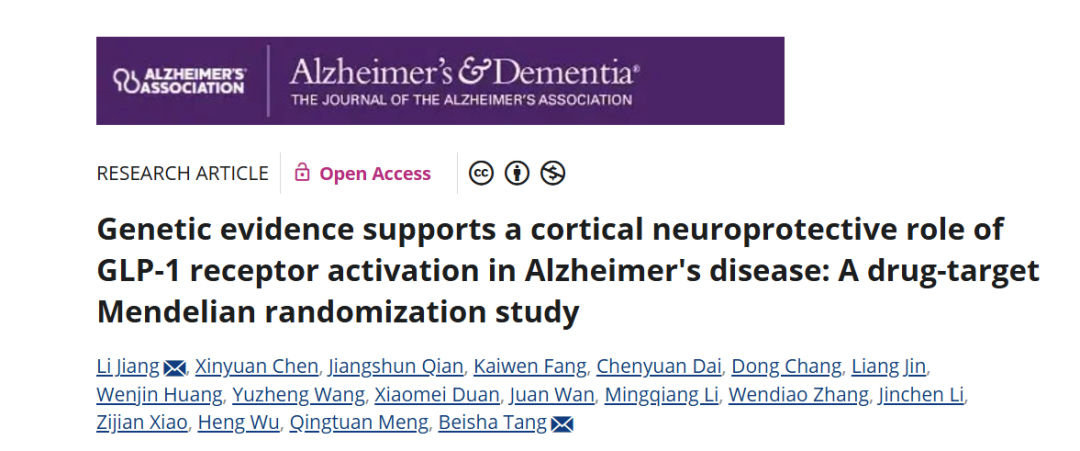 Fig. 1. Screenshot of the Published Study
Article Key Points
- Drug-target Mendelian randomization using cis-eQTLs for GLP-1R expression, not glycemic proxies
- 21,982 Alzheimer’s cases and 41,944 controls; replicated in a European Alzheimer’s biobank
- Blood GLP-1R expression and AD risk: OR 0.94 (95% CI 0.91-0.98, p=0.002)
- Cortical GLP-1R expression and AD risk: OR 0.85 (95% CI 0.75-0.96, p=0.009)
- Tissue-specific modeling: cortical PIP 0.998, blood expression excluded (PIP 0.002)
- GLP-1R expression associated with lower CSF p-tau; no association with CSF Abeta42
- Limitations: European ancestry only; cortical eQTL from 4 SNPs explaining 2.74% of variance
- Genetic evidence supports cortical neuroprotection but does not establish clinical benefit
Why Observational and Randomized Evidence Disagree
GLP-1RAs were first developed for type 2 diabetes and obesity. Their weight-lowering and cardiovascular effects have drawn attention in recent years, but evidence on cognition is not consistent: large observational studies, national medical record analyses, and target trial emulation suggest that treated patients may have a 30% to 70% lower risk of dementia or AD. Yet the ELAD trial of liraglutide in mild-to-moderate AD, and the EVOKE and EVOKE+ trials of oral semaglutide in early symptomatic AD, showed no significant cognitive benefit.
The investigators argue that the two bodies of evidence are not contradictory. One explanation is that observational studies carry indication bias and residual confounding, so the protective effect is overestimated. The other is that semaglutide, as a large peptide, may have limited blood-brain barrier penetration; even with pronounced peripheral metabolic effects, it may not achieve sufficient GLP-1R engagement in the central nervous system, which would limit any neuroprotective effect.
Design: Drug-Target Mendelian Randomization
The study did not use blood glucose or glycated haemoglobin (HbA1c) as a proxy for drug exposure. It took GLP-1R gene expression itself as the exposure and used cis expression quantitative trait loci adjacent to the GLP-1R gene as genetic instruments, mimicking the design of a randomized controlled trial to reduce confounding and reverse-causation bias inherent in observational research.
To test the validity of the selected instruments, the researchers set four positive controls: type 2 diabetes, BMI, HbA1c, and the insulin fold change (IFC, the ratio of the 2-hour post-load insulin level to the fasting level in an oral glucose tolerance test). The first three are established clinical effects of GLP-1RAs. IFC characterizes the insulin secretory response to a glucose stimulus and can capture the known effect of GLP-1R activation in promoting insulin secretion. Reproducing known effects would support the biological relevance of the instruments and, to some extent, reflect the established pharmacological actions of GLP-1RAs.
Blood Expression and Alzheimer’s Disease Risk
The study included 21,982 AD cases and 41,944 controls and replicated the analysis in the European Alzheimer’s and Dementia Biobank. Genetic instruments came from GTEx v10 whole blood (803 individuals) and MetaBrain cerebral cortex (2,439 individuals).
In the blood cis-eQTL model, GLP-1R expression was associated with a significantly lower risk of AD (OR 0.94, 95% CI 0.91-0.98, p=0.002). The replication in the European Alzheimer’s and Dementia Biobank gave OR 0.98 (95% CI 0.95-1.00, p=0.024), in the same direction. The protective effect was stronger in the cortical model (OR 0.85, 95% CI 0.75-0.96, p=0.009).
Genetically predicted GLP-1R expression was associated with higher LDL-C (beta 0.032, 95% CI 0.016-0.049, FDR-adjusted p=2.02×10⁻⁴), opposite in direction to the lipid-lowering effect seen in trials. The investigators explained this in two ways: the lipid-lowering effect of GLP-1RAs is modest overall and highly heterogeneous between agents (semaglutide lowers LDL-C appreciably, whereas liraglutide and exenatide show inconsistent effects), and more importantly, the genetic instrument reflects lifelong differences in gene expression, whereas drugs in trials are given late and for a short period, so the two cannot be equated directly.
Only Systolic Blood Pressure Survives as a Mediator
The investigators assessed 11 candidate mediators one by one and ranked them with MR-BMA. Seven variables had a relatively high marginal inclusion probability, but only BUN, SBP, and TG showed a GLP-1R to mediator to AD effect in the same direction as the protective effect; the others behaved more like suppressors than true mediators.
In the two-step multivariable analysis, only SBP retained an independent association with AD risk (cis-MRBEE: theta 1.320, 95% CI 0.307-2.333, PIP 0.840, p=0.011; cis-MVIVW: beta 0.819, 95% CI 0.451-1.187, p=1.3×10⁻⁵). BUN and TG were no longer independent after SBP was added, suggesting that their priority may derive from shared variance with SBP.
More importantly, even after simultaneous adjustment for the three highest-priority mediators, the direct protective effect of GLP-1R on AD risk remained significant (cis-MRBEE: theta -0.028, 95% CI -0.052 to -0.005, p=0.018; cis-MVIVW: beta -0.019, 95% CI -0.037 to -0.001, p=0.038).
Cortical Expression Is the Main Causal Exposure
In tissue-specific multivariable analysis, SuSiE fine mapping identified cerebral cortical GLP-1R expression as the main causal exposure (PIP 0.998, theta -0.159, 95% CI -0.216 to -0.102, p=3.69×10⁻⁸), while blood GLP-1R expression was excluded (PIP 0.002). cis-MVIVW confirmed this: cortical expression remained significant (beta -0.099, 95% CI -0.156 to -0.042, p=7.31×10⁻⁴), whereas blood expression did not (beta -0.008, 95% CI -0.018 to 0.002, p=0.105).
In other words, when blood and cortical GLP-1R expression were modeled together, the signal came mainly from the cortex rather than from blood, which the investigators describe as direct human genetic evidence for central neuroprotection.
Among AD-related endpoints, GLP-1R expression was associated with lower CSF p-tau (beta -0.052, 95% CI -0.095 to -0.009, p=0.017), suggesting that it may exert neuroprotection by inhibiting tau hyperphosphorylation. No association was found between GLP-1R expression and CSF Abeta42, offering preliminary genetic evidence that the mechanism may involve tau rather than amyloid-beta; this requires validation in larger samples.
Clinical Implications and Limitations
The study indicates that human genetic evidence supports a protective role of GLP-1R in AD risk and CSF p-tau pathology that cannot be fully explained by peripheral metabolic improvement and may involve cortical neuroprotection. This implies that future trials may need to prioritize whether a drug penetrates the blood-brain barrier effectively, and may consider intervening in the preclinical or prodromal phase of AD.
The study is limited to people of European ancestry, so generalizability is unclear. The cortical eQTL was based on only 4 SNPs in MetaBrain and explained 2.74% of variance, limiting statistical power. cis-eQTLs reflect gene expression rather than receptor number, signalling activity, or pharmacological effect itself. These results still require replication with larger brain tissue eQTL datasets and larger AD datasets.
Reference
[1] Li Jiang , Xinyuan Chen , Jiangshun Qian ,et al. Genetic evidence supports a cortical neuroprotective role of GLP-1 receptor activation in Alzheimer's disease: A drug-target Mendelian randomization study. Alzheimer's & Dementia. 2026;22:e71845.

