


Sixteen-year-old Xiaoye, a pseudonym, recently noticed that he could not see the words on the classroom blackboard clearly. At the ophthalmology department of the Second Affiliated Hospital of Zhejiang University School of Medicine (SAHZU), the receiving physician quickly referred him to neurosurgery.
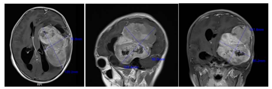
When the MRI result appeared, Dr. Xu Jinfang, chief neurosurgeon, fell silent. In more than 20 years of practice, he had seen many giant intracranial tumors, but this boy's scan still stunned him: the longest diameter was 12 cm, and the cranial cavity was almost filled.
"I have worked for many years and had never seen a brain tumor this large," Dr. Xu told MedJ, "Based on the journal literature and public reports I reviewed, excluding tumors growing outside the skull, this may be the largest tumor inside the brain."
Dr. Xu accepted the challenge. With two assistants, he removed the massive intracranial tumor completely in just over six hours. About one month after surgery, Xiaoye was discharged home, able to walk and speak normally, with no neurological deficit.
A years-long silent tumor filled the cranial space
A 12-cm brain tumor is difficult to imagine. The cranial cavity formed by the skull houses the brain, whose normal adult size is about 10 cm by 13 cm by 16 cm. By age 16, the cranial cavity is nearly adult in size.
Xiaoye's tumor almost occupied the entire intracranial space. The frontal, temporal, parietal, and occipital lobes, as well as the thalamus, were affected, and normal brain tissue had been compressed toward the skull edge and deep cranial regions. "The tumor had grown to the limit of what the intracranial space could hold."Dr. Xu said.
Yet before this visit, Xiaoye had only occasional dizziness and headache. He otherwise lived normally, studied, and exercised without obvious problems. Dr. Xu explained that the tumor was probably benign and may have existed since childhood, growing slowly over many years. The developing brain can compensate by shifting tissue away from pressure, allowing the tumor to grow silently.
If the tumor had been malignant, it would likely have grown rapidly, causing intracranial pressure to rise within a short period and making coma more likely. Brain tumors are not rare in children and adolescents. A 2022 New England Journal of Medicine review reported that brain tumors account for about 20% of all pediatric tumors, second only to leukemia, and are a leading cause of cancer-related death in children and adolescents.
Even benign intracranial tumors can have severe consequences because the skull is a closed space. As a tumor grows, less room remains for the brain. Functions such as vision, language, movement, and memory can fail depending on the area compressed. In Xiaoye's case, downward pressure affected the visual pathway, explaining his difficulty seeing the blackboard. Continued growth could have compressed the brainstem, threatening breathing, heartbeat, blood pressure, and life itself.
A skull transformed into a honeycomb
During preoperative multidisciplinary discussion, anesthesiologists were the first to raise concern. Long-term compression had kept Xiaoye's intracranial pressure high. To maintain blood flow to the brain, the body raises systemic blood pressure. But anesthetic drugs can lower blood pressure, and in a patient relying on high pressure to counter intracranial pressure, a sudden drop may cause cerebral ischemia and hypoxia.
The second major risk was bleeding. Contrast-enhanced MRI showed extremely rich blood supply to the tumor. The neurosurgical team consulted neurointerventional specialists about embolizing feeding vessels before surgery, but the tumor was supplied by multiple vessels from several directions, so the benefit was limited. The team decided that vessels would have to be sealed one by one during surgery.
On March 5, Dr. Xu led the operation with two assistant surgeons, a scrub nurse, a circulating nurse, and two anesthesiologists. Soon after the operation began, the feared problem appeared. 'As soon as we lifted the scalp, the whole skull was bleeding,' he recalled. It was not ordinary oozing but bleeding from all directions.
A normal skull is about 5 mm to 1 cm thick. Xiaoye's skull was less than 2 mm thick and had been remodeled into a honeycomb by the tumor. The tumor had long secreted vascular growth factors, drawing meningeal vessels into the skull. Tiny blood vessels drilled small holes through the bone, weaving inside and outside it. Once the scalp was opened, the whole skull surface bled.
Dr. Xu and his assistants sealed every gap on the skull surface with bone wax, controlled bleeding during craniotomy, and then sawed open the skull. It was tightly adherent to the dura and tumor. Every step, including opening the dura, brought new bleeding points that had to be addressed before proceeding.
Fast where safe, slow where vital
Dr. Xu compared the giant tumor to a large sweet potato buried in soil: removing it intact without damaging the soil requires exceptional technique. The tumor was tightly adherent to brain tissue, involved the motor cortex and thalamus, and intertwined with deep blood vessels and nerves.
Moving too slowly could extend surgery beyond 10 hours, increasing infection, massive bleeding, hemorrhagic shock, and coagulation problems. Moving too quickly could damage critical tissue, nerves, and vessels, causing disability, coma, or death.
Dr. Xu's strategy was to speed up in areas with clear visualization and easily controlled vessels, removing large tumor pieces to shorten operative time. Deeper regions near the thalamus and key vessels and nerves required much slower work, using fine microsurgical scissors under the microscope to advance fractions of a millimeter at a time. After the deepest portions were removed, the operation achieved total resection in under seven hours.
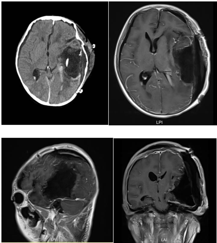
Postoperative CT and MRI showed total tumor resection with no cerebral hemorrhage or infarction. Xiaoye was observed in the intensive care unit, where his heart rate, blood pressure, consciousness, limb movement, and breathing were stable. He soon returned to the general ward.
Rounds showed no hemiplegia, aphasia, coma, severe infection, epilepsy, or other major adverse outcome. Pathology confirmed Dr. Xu's judgment: a benign meningioma. Xiaoye is now recovering at home and can walk and talk normally.
Dr. Xu said the case reinforced a key point: even a very large brain tumor should not automatically be feared. The decisive questions are whether surgeons can control intraoperative bleeding and protect normal brain function. If both can be achieved, surgery can be feasible and outcomes may be good.

