


Introduction
In hepatology, liver biopsy has long been considered the gold standard for histological diagnosis. As non-invasive technologies continue to advance, however, one question is repeatedly raised: in the era of elastography, CT, MRI, and various biomarkers, why do we still need liver biopsy?
The answer is straightforward: for some patients with complex liver disease, histological evidence remains decisive for diagnosis and treatment strategy. This is particularly true in obesity-related liver disease (MASLD/MASH), liver transplantation, focal hepatic lesions, and unexplained hepatic dysfunction. Recently, the International Obesity Society (IOS) Working Group on Steatotic Liver Disease formally published in Cell Reports Medicine the “An International Multidisciplinary Consensus Statement on Laparoscopic Liver Biopsy” — a 45-expert, six-continent collaboration. This is the first international multidisciplinary consensus on laparoscopic liver biopsy (LLB), establishing systematic standards for indications, high-risk scenarios, perioperative management, quality control, and multidisciplinary collaboration. The corresponding authors are Prof. Minghua Zheng of the First Affiliated Hospital of Wenzhou Medical University, Prof. Hua Yang of the First Affiliated Hospital of Jinan University, and Prof. James Neuberger of Queen Elizabeth Hospital Birmingham, United Kingdom.
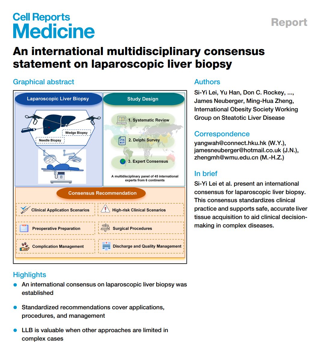 Fig. 1. Paper Title Page Summarizing LLB, Study Design, and Consensus Recommendations
Image source: Cell Reports Medicine, original extract
Why the World Needs a Consensus on Laparoscopic Liver Biopsy
Although LLB has been performed for decades, global standards have long been lacking. Centers differ substantially on:
l When to choose LLB
l How to perform preoperative evaluation
l How to obtain high-quality specimens
l How to manage complications
l How to establish quality control systems
Some centers underuse the technique, leaving complex patients without access to histological diagnosis; others apply it too broadly, increasing unnecessary anesthetic and surgical risk. A unified international standard is therefore a pressing clinical need.
Based on a systematic review and three rounds of Delphi expert surveys plus international multidisciplinary discussion, the consensus ultimately formed 46 recommendations, establishing the first global evidence-based framework for LLB (Fig. 2).
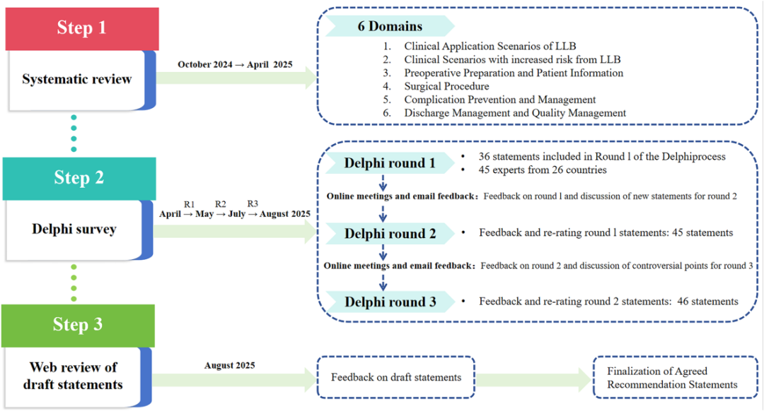 Fig. 2. Consensus Formation Process, Showing Systematic Review, Delphi Survey, Expert Feedback, and Final Consensus Development
Image source: Cell Reports Medicine, original extract
Is Liver Biopsy Still Important?
Recent years have seen enormous progress in non-invasive liver assessment. FibroScan, MRI-PDFF, MRE, CT, and various serum-based scores can already accomplish much of the screening and risk-stratification workload.
For the following scenarios, however, histological examination remains irreplaceable:
① Unexplained hepatic dysfunction
When laboratory and imaging studies cannot identify the cause, pathology remains the final diagnostic reference.
② Indeterminate hepatic nodules
Imaging cannot reliably distinguish inflammation, regenerative nodules, early hepatocellular carcinoma, or metastases.
③ Precise MASLD/MASH assessment
Defining inflammatory activity, ballooning, and fibrosis stage is essential to guide treatment decisions.
④ Post-transplant graft dysfunction
Differential diagnosis includes rejection, infection, and drug-induced injury.
⑤ Discrepancy between clinical and imaging findings
Pathology provides the definitive evidence.
In short, non-invasive testing answers “how great is the risk,” while histology answers “what the disease actually is.”
Why LLB Is the Solution for Complex Cases
Current liver biopsy approaches include:
l Percutaneous liver biopsy (PLB)
l Transjugular liver biopsy (TLB)
l Endoscopic ultrasound-guided liver biopsy (ELB)
l Laparoscopic liver biopsy (LLB)
For the vast majority of patients, percutaneous biopsy remains the first choice.
In selected complex cases, however, LLB offers distinct advantages (Fig. 3).
1. Direct visualization for precise sampling
The operator can directly inspect the liver surface, avoid major vessels and bile ducts, and improve sampling accuracy.
2. Multi-site sampling
For diseases with substantial histological heterogeneity such as MASLD, multiple-site sampling reduces sampling error.
3. Immediate hemostasis
After sampling, bleeding can be observed immediately and managed by electrocoagulation or local hemostasis.
4. Concurrent liver assessment
During bariatric/metabolic or cholecystectomy procedures, liver tissue can be obtained at the same operation, avoiding repeat anesthesia and puncture.
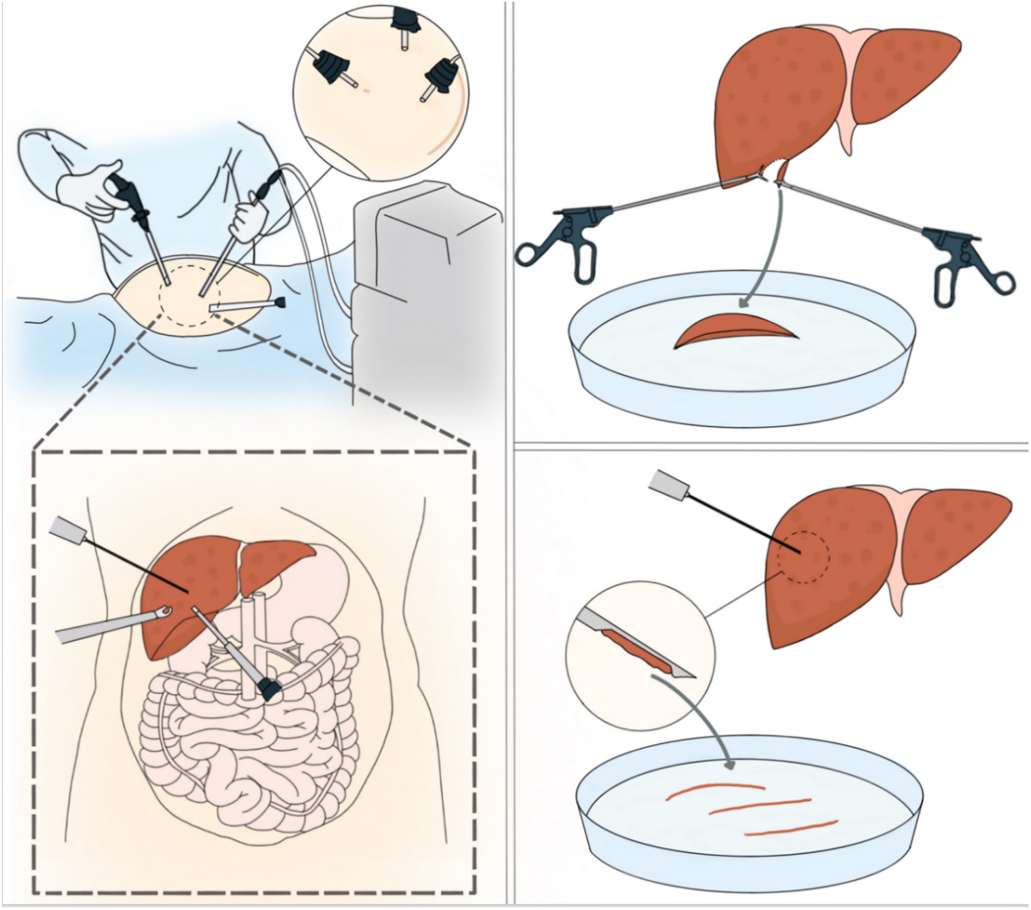 Fig. 3. Schematic of LLB Procedure, Showing Laparoscopic Direct-Vision Needle Biopsy and Wedge Biopsy.
Image source: Cell Reports Medicine, original extract
Why the Obesity and Bariatric Field Should Pay Particular Attention
This is the most practically meaningful part of the consensus.
As MASLD has become the most common chronic liver disease worldwide, large numbers of patients undergoing bariatric and metabolic surgery also have varying degrees of liver injury.
A growing body of research shows that even when imaging suggests only fatty liver, some patients already harbor significant inflammation or even advanced fibrosis. As a result, intraoperative opportunistic liver biopsy has attracted increasing attention.
The consensus explicitly notes that for patients undergoing bariatric and metabolic surgery, intraoperative LLB can: accurately assess fibrosis stage, identify occult MASH, generate real-world histological evidence, and prevent missing advanced liver disease after surgery. The implication is clear: bariatric surgery is not only an opportunity to treat obesity, but may also become an important window for precisely assessing liver health.
Who Should Be Prioritized for LLB — and Who Requires Extreme Caution
The consensus summarizes six core indications (Fig. 4):
① Conventional biopsy has failed or is not feasible
Including failed percutaneous biopsy, contraindicated or limited transjugular biopsy, and inadequate EUS sampling.
② Lesions in special anatomic locations
For example, hilar region, caudate lobe, and subdiaphragmatic lesions.
③ Diffuse liver disease requiring multi-site sampling
Including MASLD/MASH, hepatic fibrosis, and inflammatory liver disease.
④ Special coagulation disorders
LLB may serve as an important complement to transjugular liver biopsy after thorough evaluation.
⑤ Opportunistic biopsy during bariatric/metabolic surgery
Liver tissue obtained concurrently with the bariatric procedure.
⑥ Special complex patients
Including evaluation of living liver donors, post-transplant patients, unexplained ascites, NCPF/IPH.
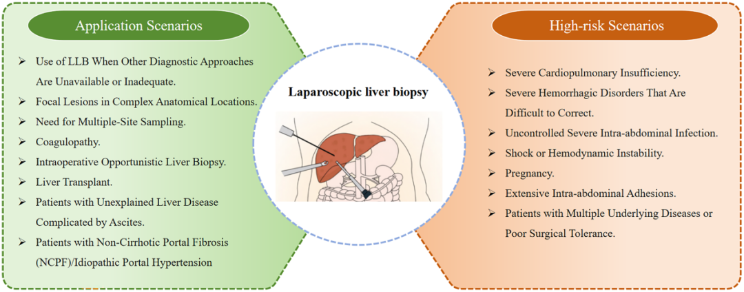 Fig. 4. Indications for LLB and High-Risk Scenarios: Clinical Scenarios Where LLB May Be Considered (Left) and Situations Requiring Careful Evaluation (Right)
Image source: Cell Reports Medicine, original extract
Seven Populations Requiring Extreme Caution with LLB
Laparoscopy requires pneumoperitoneum and general anesthesia, which impose systemic demands on the patient. The consensus specifies the following high-risk, restricted-use populations:
1. ASA IV-V with severe cardiopulmonary dysfunction unable to tolerate pneumoperitoneum or anesthesia;
2. Refractory severe coagulopathy — transjugular liver biopsy preferred;
3. Uncontrolled intra-abdominal active infection or sepsis;
4. Shock or hemodynamically unstable patients;
5. Late pregnancy (≥28 weeks), when the uterus occupies the abdominal cavity and limits operative space; early-to-mid pregnancy may proceed only after MDT evaluation when benefit clearly outweighs risk;
6. Extensive intra-abdominal adhesions, with very high risk of visceral injury during dissection;
7. Multiple comorbidities stacked together, with very poor surgical tolerance.
The above are not absolute contraindications, but must be evaluated jointly by hepatology, surgery, anesthesia, and ICU teams, with strict control of operative indications.
A Complete Preoperative–Intraoperative–Postoperative Workflow
What most distinguishes this consensus from prior literature is that it answers not only “whether to do it” but also “how to do it correctly.”
The content covers:
1. Preoperative evaluation
Imaging time window, coagulation assessment, anticoagulant management, anesthesia risk evaluation.
2. Intraoperative procedure
Pneumoperitoneum pressure standards, choice of sampling site, required tissue length, intraoperative ultrasound use.
3. Postoperative management
Discharge criteria, complication monitoring, follow-up system.
4. Quality control
Specimen quality requirements, physician training standards, MDT management system.
Together, these mark LLB’s formal entry into the era of standardization.
Three Breakthrough Values of the Consensus
First, the consensus clarifies the appropriate boundary of LLB. It is not a replacement for routine biopsy methods, but an important complementary tool for complex or high-risk scenarios.
Second, the consensus expands LLB from a single surgical procedure into a complete workflow — covering preoperative evaluation, intraoperative sampling, complication management, discharge criteria, follow-up planning, and quality control — helping move the technique from experience-based to standardized practice.
Third, the consensus emphasizes multidisciplinary collaboration. For patients with complex liver disease or high procedural risk, the value of histological diagnosis must be weighed together with procedural risk. The MDT model helps improve decision quality, reduce unnecessary risk, and enhance diagnostic efficiency.
Conclusion
From a histological perspective, liver biopsy remains an irreplaceable tool in modern hepatology. The value of LLB does not lie in being “more advanced,” but in helping complex patients obtain high-quality histological evidence more safely and precisely. The release of this international consensus fills the long-standing gap in unified standards for laparoscopic liver biopsy worldwide, and provides a new evidence base for precision care in obesity-related liver disease, bariatric/metabolic surgery, and complex liver disease. As the disease burden of MASLD and MASH continues to grow, LLB is expected to play an increasingly important role at the intersection of bariatric medicine and hepatology.
Reference
[1] Lei SY, Han Y, Rockey DC, Alswat K, Wong VW, Jumaev N, Toro-Huamanchumo CJ, Taha S, El-Kassas M, George J, Muthiah M, Ghanem O, Yoneda M, Hang DV, Sebastiani G, Papatheodoridis G, Abu-Abeid A, Robertson AG, Kim SH, Weiner R, Mahawar K, Shabbir A, Kermansaravi M, Rajan R, Seki Y, Taskin HE, Shikora S, Oliveros E, Parmar C, Cheng Q, Thaher O, Ruiz-Úcar E, Oviedo R, Ribeiro R, Musella M, Ramos AC, Ryu SW, Abdelbaki T, Lee WJ, Chan MP, Garcia Flores JE, Skinovsky J, Madkhali A, Weiner S, Tacke F, Yang W, Neuberger J, Zheng MH; International Obesity Society Working Group on Steatotic Liver Disease. An international multidisciplinary consensus statement on laparoscopic liver biopsy. Cell Rep Med. 2026 Jun 16;7(6):102831. doi: 10.1016/j.xcrm.2026.102831. Epub 2026 May 22. PMID: 42173098.

