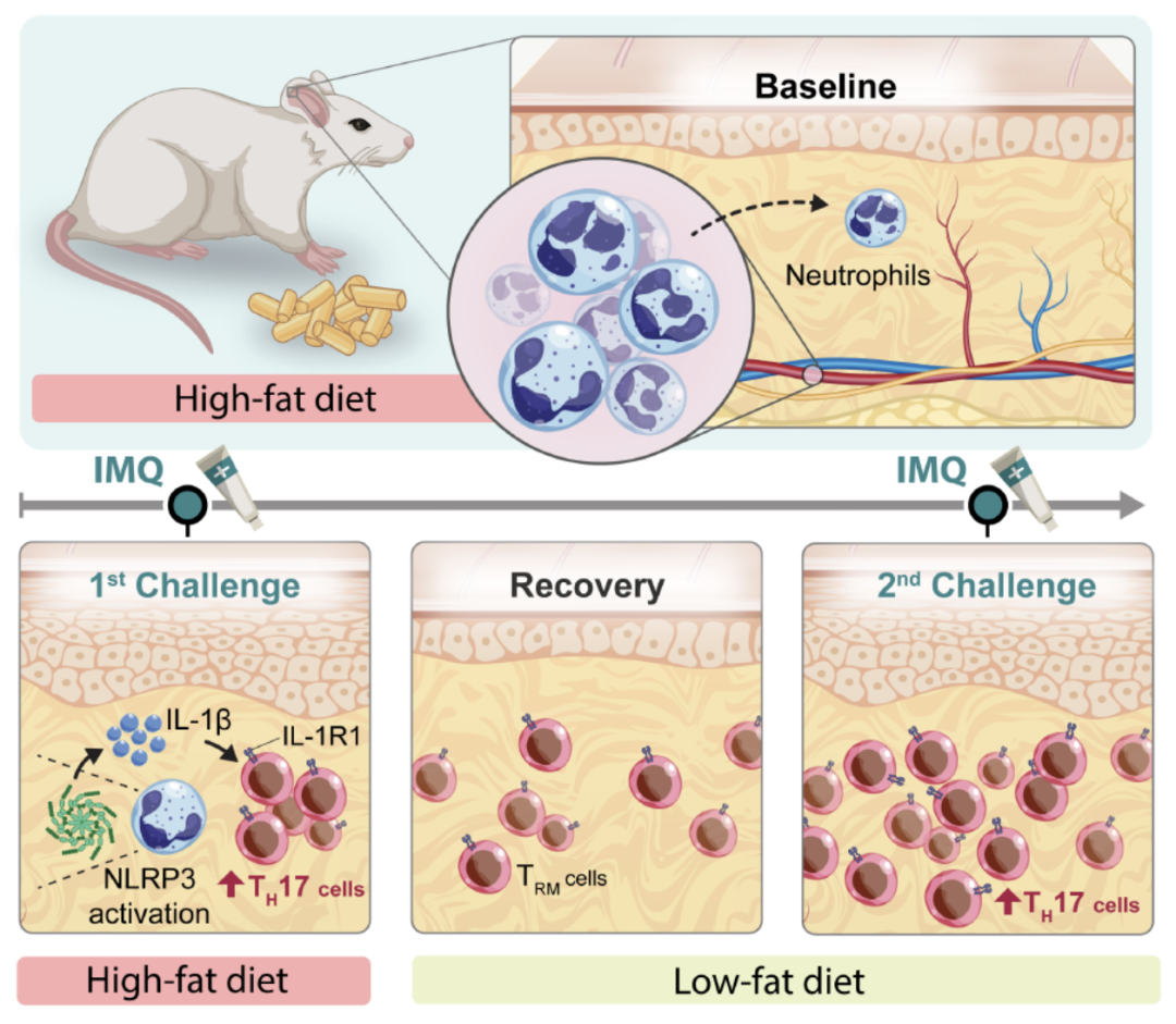
Obesity alters immune function in infection, autoimmunity, and cancer. Its effects are particularly evident in the skin, where obesity can aggravate inflammation and influence treatment responses. Chronic obesity and obesogenic diets consumed for weeks or months are known to remodel cutaneous immunity, but these prolonged interventions also produce cumulative metabolic, endocrine, microbial, and tissue changes across multiple organ systems. Whether dietary changes lasting only days can alter induced inflammation before such systemic remodeling becomes established, and whether they leave durable tissue inflammatory memory, has remained unclear.
A study led by first author Dr. Zewen Jiang of the University of California, San Francisco, was published in Nature on October 28, 2026, under the title “Diet at disease onset determines tissue inflammatory memory”. It reports that several days of high-fat diet (HFD) exposure can persistently reshape tissue immune states in mice without causing obesity. The findings implicate the dietary state at disease onset, rather than accumulated adiposity alone, in both acute amplification of inflammation and longer-term changes in local immune responsiveness.
Article Key Points
- The study tested whether days of dietary change alter inflammation before systemic remodeling develops
- Four days of high-fat feeding amplified imiquimod-induced ear inflammation without causing obesity in mice
- Obese mice switched to a low-fat diet for four days had substantially milder inflammation
- Rechallenge weeks after dietary reversal and clinical resolution triggered an exaggerated response in the same ear
- Neutrophil depletion, Nlrp3 deletion, or NLRP3 inhibition reduced high-fat diet-induced inflammatory amplification
- The neutrophil-NLRP3-IL-1β axis activated pathogenic IL-1R1+ CD4+ TH17 cells
- Tissue-resident memory T cells persisted in previously inflamed skin after the acute response subsided
- Brief high-fat feeding also enhanced pulmonary TH17 inflammation through neutrophil- and IL-1R1-dependent mechanisms
- The findings are preclinical and do not establish equivalent effects of short-term dietary changes in humans
The investigators initially fed mice either low-fat or high-fat chow and applied imiquimod, a cream that induces skin inflammation, to one ear of each animal. Obese mice receiving habitual high-fat feeding developed severe responses, whereas mice fed low-fat chow had mild responses. Dr. Jiang said the team wanted to determine whether excess weight itself or the diet was driving the stronger inflammatory response.
To separate these factors, mice raised on low-fat chow were switched to high-fat food. Imiquimod was applied to one ear after 3 days, within a 4-day dietary exposure that was insufficient to increase body weight. Despite remaining at normal weight, these mice developed severe ear inflammation resembling that seen in obese mice. Conversely, obese mice switched to a low-fat diet for 4 days showed markedly reduced inflammation.
These experiments addressed the immediate effect of diet independently of established obesity. The investigators emphasized that long-term dietary habits remain important, while arguing that their findings reveal a more immediate influence of diet on immune responses. Their comments extended this concept to meals throughout the day, although the experiment tested defined dietary exposures in mice rather than individual meals in humans.
The team then examined whether the effect persisted beyond the initial exposure. Normal-weight mice were returned to a low-fat diet and their inflammation was allowed to resolve. Several weeks later, imiquimod was applied again to the same ear that had previously become inflamed. The rechallenge produced an exaggerated immune response, indicating that the tissue remained primed after both dietary reversal and clinical resolution of the initial lesion.
The results therefore linked a brief physiological state at disease onset with a durable change in subsequent local responses. The investigators propose that short-lived states may become incorporated into local immune memory when they coincide with tissue inflammation.
Mechanistically, high-fat feeding rapidly recruited neutrophils into the skin. Neutrophils in stimulated skin expressed high levels of Nlrp3 and Il1b. Neutrophil depletion, genetic deletion of Nlrp3, and pharmacological inhibition of NLRP3 each reduced the inflammatory amplification induced by HFD.
These findings identified a neutrophil-NLRP3-IL-1β signaling axis that activated pathogenic IL-1R1+ CD4+ TH17 cells. As inflammation subsided, tissue-resident memory T cells remained in the affected ear. Together, early innate immune amplification and persistent tissue-resident memory cells left the skin more readily activated by a subsequent stimulus. Mice exposed to HFD at disease onset consequently developed excessive inflammation on recurrence despite returning to and remaining on a low-fat diet.
A related effect was observed beyond the skin. Several days of high-fat feeding significantly enhanced TH17 inflammation in the lung through mechanisms dependent on neutrophils and IL-1R1. Across these experiments, dietary state during a narrow time window durably reshaped disease trajectories in the mouse models studied.