


A 57-year-old male patient with MSI-H, TMB-H intrahepatic cholangiocarcinoma following palliative surgery experienced multiple episodes of new hepatic lesions suggestive of disease progression during first-line and second-line therapy. These lesions were ultimately confirmed by biopsy to be inflammatory nodules (immunotherapy pseudoprogression), thereby preventing an erroneous change in treatment strategy. Continued treatment with the original regimen resulted in sustained disease stabilization.
1. Core Clinical Contradiction and Outcome
The central dilemma was: in the setting of immunotherapy, when imaging reveals "new lesions" (enlarging or de novo hepatic nodules), how can true progression be distinguished from pseudoprogression? Misclassifying the findings as true progression would lead to premature abandonment of an effective regimen. Ultimately, biopsy of the enlarging lesion revealed "inflammatory cell infiltration with no tumor cells," confirming immunotherapy pseudoprogression. Continuation of the original regimen resulted in lesion regression and sustained long-term disease control.
2. Clinical Implications
For patients with cholangiocarcinoma — especially those with MSI-H/TMB-H tumors who represent an immunotherapy-favored population — isolated, localized "radiographic progression" following immunotherapy should not be taken as definitive progression. When feasible, prompt biopsy to clarify the pathological nature of suspicious lesions is the critical tool for differentiating true progression from pseudoprogression, thus avoiding premature termination of effective treatment.
1. Case Overview: Baseline Characteristics and Core Challenges
Baseline Status: A 57-year-old male underwent "radical left intrahepatic cholangiocarcinoma resection (left hemihepatectomy + cholecystectomy + lymph node dissection + hepatic artery reconstruction + hepaticojejunostomy)" in June 2024.
Postoperative Pathology: Intrahepatic cholangiocarcinoma, moderately to poorly differentiated, with vascular tumor thrombus and perineural invasion; lymph node metastasis (11/17 nodes positive).
Genomic Profiling: TMB: 52.95 muts/Mb; MSI: MSI-H. One month after surgery, prior to initiating systemic therapy, a biopsy of the left supraclavicular lymph node (which had been present on preoperative baseline imaging) confirmed metastatic cholangiocarcinoma, establishing the final stage as pT2N1M1, stage IV.
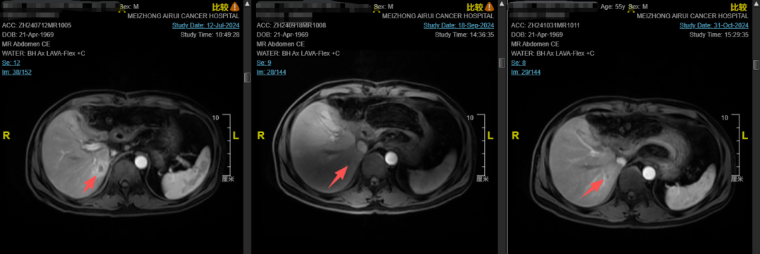 Fig. 1. Liver MRI After First-Line Treatment
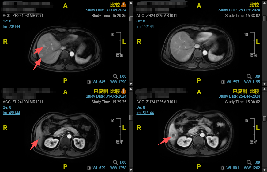
Fig. 2. Liver MRI After Second-Line Treatment
Core Challenges:
First-line therapy with lenvatinib plus pembrolizumab for 6 cycles was followed by imaging that revealed new hepatic nodules suggestive of multiple metastases, with an assessment of progressive disease (PD).
After 2 cycles of second-line therapy with regorafenib plus gemcitabine plus cisplatin plus cadonilimab, imaging again showed that while some hepatic lesions had regressed, a lesion in the inferior segment of the right posterior lobe had enlarged significantly compared with prior scans.
Faced with this "mixed response," how should one accurately determine whether the treatment is effective? Should the regimen be changed?
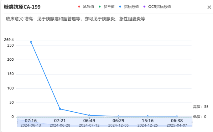
Fig. 3. Serial Serum CA19-9 Levels Following Surgery
2. Decision Discussion: MDT Analysis and Regimen Deliberation
MDT Team Composition
The multidisciplinary team comprised specialists from Medical Oncology, Hepatobiliary Surgery, Interventional Radiology, Diagnostic Imaging, and Pathology.
Treatment Pathway Reasoning and Risk-Benefit Analysis
After 2 cycles of second-line therapy, the efficacy assessment indicated a mixed response — some lesions had regressed while others had enlarged — reflecting a mixed response in the setting of tumor heterogeneity. The MDT focused their discussion on determining the true nature of the enlarging lesion.
Multidisciplinary Perspectives
Option A
Classify as true progression and switch the treatment regimen immediately.
According to the conventional RECIST criteria for solid tumor response evaluation, enlarging lesions or new nodules can directly indicate PD, warranting immediate discontinuation of the current regimen and initiation of post-second-line salvage therapy.
Advantage: Prompt cessation of ineffective treatment, avoiding continued disease progression.
Drawback: The patient has an MSI-H/TMB-H tumor — an immunotherapy-favored profile associated with high likelihood of benefit. The majority of lesions had already demonstrated treatment response. Reliance solely on imaging for efficacy assessment carries a high risk of misjudgment and would prematurely terminate an effective immunotherapy regimen. Furthermore, salvage options for advanced ICC are limited, and switching regimens could substantially curtail the patient’s survival benefit.
Option B
Classify as immunotherapy pseudoprogression and continue the current regimen.
Given the patien’s history of immunotherapy exposure, an overall reduction in tumor burden, and the atypical progression pattern characterized by an isolated enlarging lesion, the likelihood of immunotherapy pseudoprogression was considered high.
Advantage: If correctly identified, sustained treatment with the effective regimen would control the tumor and maximize the long-term benefit of immunotherapy.
Risk: If the lesion represents true tumor progression, continued treatment with an ineffective regimen could lead to rapid disease progression and delay the optimal window for alternative therapy.
Option C
Perform a biopsy of the suspicious lesion to obtain pathological confirmation and guide individualized decision-making.
The enlarging hepatic lesion was targeted for core needle biopsy to establish the pathological gold-standard diagnosis for the lesion and guide subsequent treatment.
Advantage: Provides definitive differentiation between true progression and immune-mediated inflammatory pseudoprogression, enabling precise decision-making without the risk of subjective misjudgment.
Risk: The biopsy procedure carries inherent risks of minor trauma, bleeding, and infection. However, in this case the puncture trajectory was anatomically safe and the overall risk was considered manageable.
3. Treatment Execution: MDT-Guided Regimen Evolution
Treatment Roadmap
Left hemihepatectomy → First-line: Lenvatinib + Pembrolizumab, 6 cycles (PD on imaging)
→ Second-line quadruple regimen: Regorafenib + Gemcitabine + Cisplatin + Cadonilimab, 2 cycles (mixed hepatic response: partial lesion regression with isolated lesion enlargement)
→ Core needle biopsy of suspicious hepatic lesion: extensive inflammatory cell infiltration, no tumor cells identified; pseudoprogression confirmed
→ Regorafenib + Cadonilimab maintained; GP chemotherapy discontinued
→ Due to severe allergic reaction and infusion-related reactions to cadonilimab, switched to Nivolumab + Ipilimumab dual immunotherapy combined with regorafenib
→ Multiple subsequent imaging assessments: stable disease (SD)
→ Current regimen: Nivolumab monotherapy combined with reduced-dose regorafenib as maintenance therapy.
In-Depth Analysis of Key Decision Nodes
The pivotal decision node in this case was the biopsy of the enlarging hepatic lesion. In the era of immunotherapy, new or enlarging lesions on imaging are not synonymous with true progression. The patient had an MSI-H/TMB-H "hot tumor," and the development of an isolated, atypical radiographic "progression" on immunotherapy was highly suggestive of pseudoprogression.
Timely biopsy confirmed the lesion to be an inflammatory nodule (immunotherapy-related reaction), thereby validating the diagnosis of pseudoprogression.
Managing Unexpected Events and Adjustments
Identification and management of pseudoprogression: When imaging showed an isolated enlarging lesion against a background of overall disease improvement, the MDT did not blindly label the case as PD. Instead, they actively pursued biopsy. The pathological result served as the definitive anchor, sparing the patient from an unnecessary regimen switch and enabling continued benefit from immunotherapy.
Management of immune-related adverse events (irAEs): The patient subsequently developed cadonilimab-related cutaneous allergic reactions (rash with pruritus) and multiple infusion-related reactions (hypotension, syncope). After a safety re-evaluation, the regimen was changed to nivolumab plus ipilimumab dual immunotherapy, with manageable rash and uneventful infusions thereafter.
4. Outcome Assessment and Follow-Up Strategy
Short-Term Efficacy
Following confirmation of pseudoprogression and optimization of the treatment regimen, the previously enlarging hepatic lesion showed no residual viable tumor. Pre-existing metastatic lesions continued to shrink, with some achieving complete regression. No new distant metastases have emerged throughout the entire course. Multiple serial imaging assessments have consistently demonstrated SD, confirming effective tumor control.
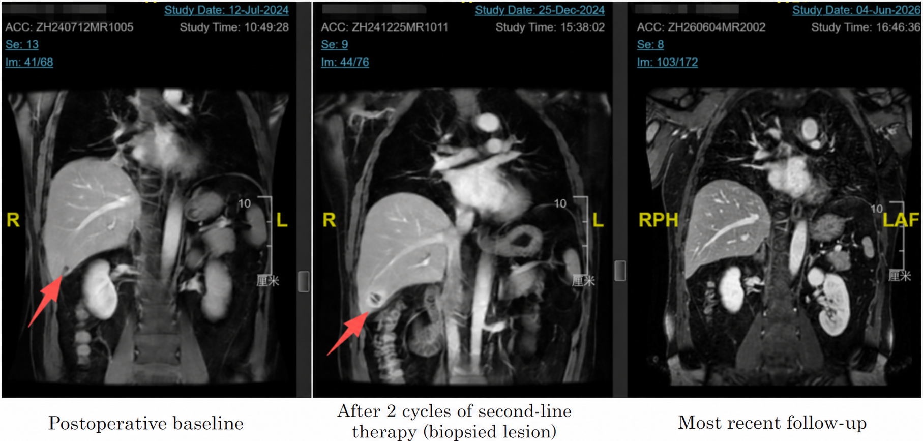
Fig.4.Postoperative Baseline,After 2 Cycles of Second‑Line Therapy (at the Punctured Lesion),Most Recent Follow‑Up Assessment
Survival and Functional Status
Since the adjustment of second-line therapy, the patient has remained in sustained SD through the most recent assessment. Throughout the postoperative treatment period, the patient has maintained an excellent performance status (PS score of 0). During the quadruple-regimen phase, transient hematuria, hypertension, and rash occurred but resolved with symptomatic management, with no grade ≥ 3 severe adverse events. After pseudoprogression was confirmed, quality of life improved further during targeted-immunotherapy, and the patient has been able to engage in physical exercise and travel.
Subsequent Treatment and Follow-Up Plan
Current treatment: Nivolumab 480 mg Q4W as maintenance; regorafenib reduced to 40 mg/day.
Surveillance plan: Upper abdominal MRI and tumor marker assessment every 2–3 months, with focused monitoring of hepatic lesions and cervical lymph node status.
Relapse contingency: Should suspicious progressive lesions reappear, the MDT will again prioritize biopsy to distinguish true progression from pseudoprogression. Subsequent treatment will be tailored based on repeat biopsy pathology and genomic testing results.
5. Key Insights from This Case
Clinical Reasoning Insights
In the immunotherapy era, clinicians must remain vigilant for pseudoprogression. For patients receiving immune checkpoint inhibitors (ICIs) — particularly those in immunotherapy-favored populations such as MSI-H/TMB-H — the appearance of new or enlarging lesions on imaging does not equate to treatment failure. A comprehensive evaluation incorporating overall tumor burden dynamics and lesion imaging characteristics (e.g., margins, perilesional edema) is essential.
Biopsy is the gold standard for distinguishing true progression from pseudoprogression. When the imaging diagnosis is equivocal and the lesion is safely accessible for biopsy, pathological evidence should be actively pursued. In this case, a single timely biopsy averted the erroneous discontinuation of an effective immunotherapy regimen and directly altered the patient’s prognosis. In retrospect, the "new hepatic nodules" observed after 6 cycles of first-line lenvatinib plus pembrolizumab were very likely also pseudoprogression. Had the same biopsy-driven approach been employed at that juncture, the nature of those lesions might have been clarified earlier, potentially obviating the need for subsequent quadruple therapy and its associated toxicities.
MDT collaboration is indispensable in complex decision-making. Diagnostic Imaging contributed insights into atypical radiographic features, Medical Oncology analyzed the probability of immunotherapy benefit, and Pathology guided the biopsy procedure and diagnosis. This multidisciplinary collaboration culminated in the correct decision: "biopsy for clarity, then continue treatment."
Implications for Clinical Practice
Pseudoprogression is a phenomenon frequently observed in clinical practice following immunotherapy. It refers to an unconventional clinical response pattern in which initial radiographic enlargement or the appearance of new lesions during ICI therapy is followed by subsequent lesion shrinkage or stabilization without a change in treatment regimen [1].
It is characterized by initial radiographic progression followed by subsequent tumor regression or stabilization without regimen modification, confirmed through repeat imaging. The pathological hallmark: in contrast to true progression (tumor cell proliferation), biopsy of enlarging pseudoprogressive lesions reveals predominantly T-lymphocyte and other inflammatory cell infiltration, necrosis, or edema, accompanied by an actual reduction in tumor burden, rather than proliferation of malignant cells.
In general, clinically common pseudoprogression predominantly manifests as lesion enlargement; the emergence of multiple new lesions is less typical, particularly for MSI-H tumors, which are highly sensitive to immunotherapy. Inflammatory pseudotumor represents a special subtype of pseudoprogression that is uncommon in clinical practice. The literature reports an incidence of approximately 2%–5% for such pseudoprogression/inflammatory pseudotumor in solid tumors such as non-small cell lung cancer (NSCLC), typically occurring early in the course of immunotherapy, though delayed presentations have also been reported [2].
At present, pathological biopsy remains the gold standard for diagnosing pseudoprogression, although clinical practice often relies on a composite assessment incorporating dynamic iRECIST evaluation, ctDNA monitoring, and clinical symptom dissociation [3,4]. When discrimination is challenging or involves critical organ function, MDT evaluation is recommended.
For patients with MSI-H solid tumors who develop recurrent new lesions following immunotherapy, the possibility of inflammatory pseudotumor should be considered. In suspected cases, it is advisable to continue treatment across radiographic "progression" for observation, provided patient safety is ensured. When feasible, proactive and safe biopsy to clarify the true nature of enlarging lesions (tumor vs. inflammation) is the critical step in avoiding premature termination of effective therapy and achieving truly individualized precision treatment. This case provides an important reference for clinical decision-making in similar scenarios.
Expert Commentary

Prof. Li Bai
Chair, Academic Committee; Chair, Ethics Committee, Beijing Arion Cancer Hospital
Member, International Committee on Challenging and Rare Tumors
Formerly: Chief Physician & Professor, Department of Medical Oncology, First Medical Center, Chinese PLA General Hospital
This is a relatively rare case of MSI-H cholangiocarcinoma. The core clinical challenge here was: during immunotherapy, imaging revealed enlarging hepatic lesions and new nodules. When the radiographic findings did not fully conform to the typical appearance of metastatic disease, yet metastasis could not be entirely excluded, the team raised the suspicion of pseudoprogression and elected to perform a confirmatory biopsy — a decision that is highly commendable and worth recommending.
Immunotherapy should not blindly adhere to the conventional RECIST imaging-based response criteria. The key question is how to accurately differentiate true tumor progression from pseudoprogression following immunotherapy. Pathological biopsy is the gold standard. However, before proceeding to biopsy, one must first evaluate whether there has been clinical symptomatic improvement, changes in laboratory parameters, and imaging characteristics that indicate tumor growth or regression — not every increase in tumor volume warrants a biopsy, especially given the low overall incidence of pseudoprogression.
Clinically, typical pseudoprogression predominantly presents as enlargement of a single lesion; multiple new lesions are relatively uncommon. This patient, by contrast, presented with multiple suspicious de novo hepatic inflammatory nodules — a rare presentation of immune-related inflammatory pseudotumor, with a very low incidence, more commonly seen early in the course of immunotherapy and highly susceptible to misdiagnosis as widespread progression. Similar cases are rare and carry significant reference value: the MSI-H/TMB-H subtype of ICC itself has a low incidence, and the presentation of immunotherapy-related multifocal inflammatory pseudotumor-type pseudoprogression — distinct from the conventional single-lesion pattern — supplements the clinical data for this unusual presentation.

Prof. Min Sun
University of Pittsburgh Medical Center (UPMC)
This is a case report of exceptional educational value. In the rare MSI-H/TMB-H cholangiocarcinoma subtype, the authors resisted the conventional RECIST reflex — "new lesion = PD = switch regimen" — and instead used biopsy pathology as the gold standard to confirm an immunotherapy-related reaction. The decision-making chain is clear and reproducible, and the management of irAEs was robust and appropriate.
Particularly noteworthy is that the authors have accurately distinguished between classical pseudoprogression and the rarer inflammatory pseudotumor/sarcoid-like reaction subtype, while also incorporating ctDNA into the comprehensive assessment framework for true versus pseudoprogression. These two points reflect a sophisticated grasp of the complexity of response evaluation in the immunotherapy era and elevate the mechanistic interpretation and clinical guidance of this article considerably above typical case reports.
References
1. Fountoukidis G, Tina E, Gothlin-Eremo A, Ullenhag G, Valachis A. Optimizing the clinical assessment of pseudoprogression in patients with solid tumors treated with checkpoint inhibitors: a systematic literature review and meta-analysis of associated features. Cancer Immunol Immunother. 2026;75(6):170.
2. Fountoukidis G, Tina E, Gothlin-Eremo A, Ullenhag G, Valachis A. Optimizing the clinical assessment of pseudoprogression in patients with solid tumors treated with checkpoint inhibitors: a systematic literature review and meta-analysis of associated features. Cancer Immunol Immunother. 2026;75(6):170.
3. Borcoman E, Kanjanapan Y, Champiat S, et al. Novel patterns of response under immunotherapy. Ann Oncol. 2019;30(3):385-396.
4. Persigehl T, Lennartz S, Schwartz LH. iRECIST: how to do it. Cancer Imaging. 2020;20(1):2.

