


Article Key Points:
- At the 2026 ESC Congress, Prof. Sabri Soussi of the University of Toronto Health Network proposed redefining cardiogenic shock as “Cardiogenic Shock 2.0,” moving beyond a single-marker diagnostic approach.
- Serum lactate elevation may reflect catecholamine stimulation, systemic inflammation, ischemia-reperfusion injury, or impaired hepatic and renal clearance, rather than tissue hypoperfusion alone.
- Central venous oxygen saturation (ScvO2) and left ventricular outflow tract velocity-time integral (LVOT-VTI) can detect hemodynamic deterioration earlier than lactate or organ-injury markers.
- Cardiogenic shock comprises distinct clinical phenotypes—AMI-CS, HF-CS, secondary, and post-procedural—and four biological subtypes identified via the ShockCO-OP platform.
- The inflammatory and cardiopathic biological subtypes were associated with significantly higher 28-day mortality, independent of SCAI shock staging, with Harrell’s C-index improving by 4%–6%.
In acute cardiovascular care, cardiogenic shock (CS) remains a leading cause of death. The traditional definition treats tissue hypoperfusion as the diagnostic gold standard; however, by the time overt hypoperfusion becomes clinically apparent, patients have often progressed to an irreversible stage of multi-organ injury. At the 2026 European Society of Cardiology (ESC) Congress, Prof. Sabri Soussi of the University of Toronto Health Network delivered a lecture titled “Cardiogenic Shock 2.0: Toward a More Precise and Clinically Useful Definition,” offering a critical reassessment of this long-standing clinical paradigm. Prof. Soussi argued that a definition targeting “early shock” is needed, rather than one that defines only “established shock.”
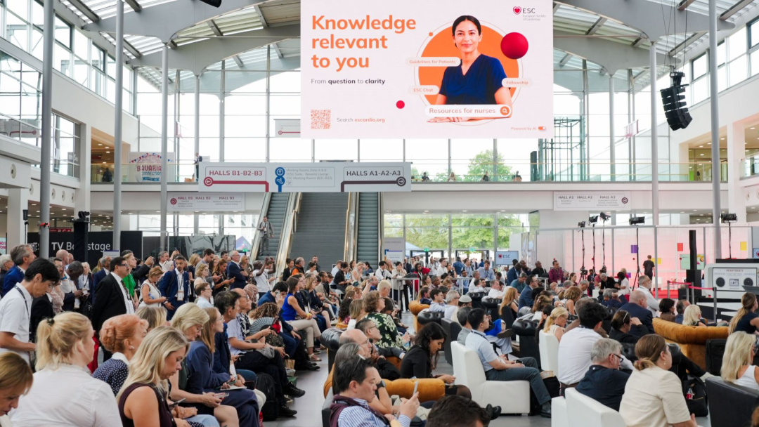 Behind-the-scenes Moments from the Venue
Rethinking Traditional Markers: Lactate Is Not a Synonym for Tissue Hypoxia
Serum lactate has long served as a key biomarker for clinicians to rapidly assess the severity of CS. However, Prof. Soussi emphasized that the pathophysiology of lactate elevation is far more complex than previously recognized. Under shock conditions, lactate levels may rise not only because of tissue hypoperfusion but also because of endogenous or exogenous catecholamine stimulation, systemic inflammatory responses, ischemia-reperfusion injury, and impaired clearance due to hepatic or renal dysfunction. Continuing to treat lactate as the gold standard—or even the primary criterion—for diagnosing CS risks misleading clinical decision-making and may lead to delayed or excessive intervention.
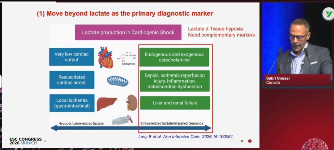 Fig. 1. Lactate Generation is Influenced by Hypoperfusion, Stress, Inflammation, and Impaired Clearance; Its Elevation is not Equivalent to Tissue Hypoxia
Gaining a Therapeutic Window: Capturing the Transition from Compensation to Decompensation
Using the metaphor of the Titanic, Prof. Soussi underscored the urgency of moving the diagnostic threshold earlier. The current revised Society for Cardiovascular Angiography and Interventions (SCAI) shock staging system defines hypoperfusion as classic shock (Stage C), but Prof. Soussi argued that the transition from “Beginning” (Stage B) to “Classic” (Stage C)—the period when “the ship has just struck the iceberg but has not yet sunk”—is where clinical attention should focus.
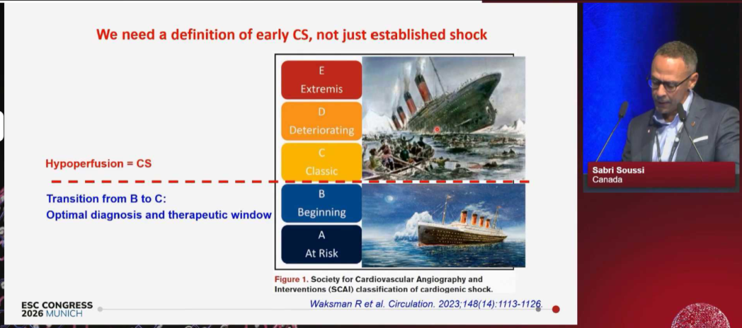 Fig. 2. The SCAI Staging Model with the Titanic Metaphor Emphasizes Identifying the “therapeutic window” of the B-to-C Transition
Drawing on experimental evidence from a porcine cardiogenic shock model and the recommendations of the 2025 European Society of Intensive Care Medicine (ESICM) guidelines, Prof. Soussi noted that central venous oxygen saturation (ScvO2) and left ventricular outflow tract velocity-time integral (LVOT-VTI) form a reliable assessment system for capturing this therapeutic window. The data showed that within 2 hours after MI-microembolization, the cardiac index and pulmonary artery pressure had already changed significantly, while serum lactate and creatinine had not yet differed notably, and only mixed ScvO2 had declined significantly (Fig. 3).
In addition, focused cardiac ultrasound (FOCUS) measurement of LVOT-VTI can provide clinicians with immediate hemodynamic information and improve risk stratification. The 2025 ESICM guidelines also recommend continuous ScvO2 monitoring in patients with shock.
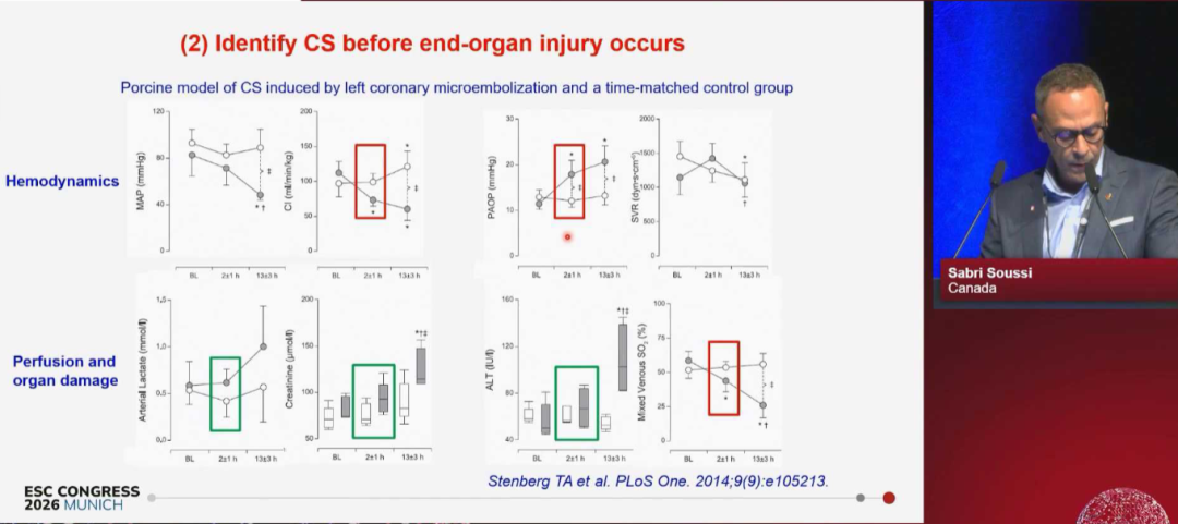 Fig. 3. Experiments Show that Hemodynamic Parameters and ScvO2 Change before Lactate and Organ-Injury Markers
From Syndrome to Heterogeneous Spectrum: Clinical and Biological Subtypes
CS is not a single disease but a highly heterogeneous syndrome with diverse etiologies and pathophysiologies. Prof. Soussi emphasized that the new definition must incorporate distinct clinical phenotypes: from an etiological perspective, acute myocardial infarction-related cardiogenic shock (AMI-CS), heart failure-related cardiogenic shock (HF-CS), secondary shock (e.g., arrhythmias, pericardial disease), and post-procedural shock each have markedly different treatment strategies and prognoses.
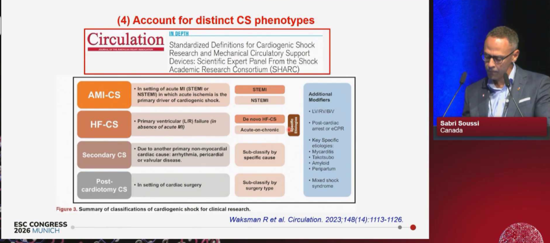 Fig. 4. Clinical Phenotype Classification of CS Based on Etiology
A more forward-looking advance lies in the identification of biological subtypes. In a study published on the ShockCO-OP platform, Prof. Soussi and colleagues performed unsupervised clustering analysis of biomarkers from 1116 CS patients and identified four biological subtypes with independent prognostic value: “Adaptive,” “Non-inflammatory,” “Cardiopathic,” and “Inflammatory” (Fig. 5). Among these, the inflammatory and cardiopathic subtypes were associated with significantly higher 28-day mortality—an association independent of SCAI shock staging. This suggests that clinical staging alone may not be sufficient to accurately predict patient outcomes, and incorporating biological phenotypes into the definition can substantially improve the accuracy of risk stratification (Harrell’s C-index improved by 4%–6%).
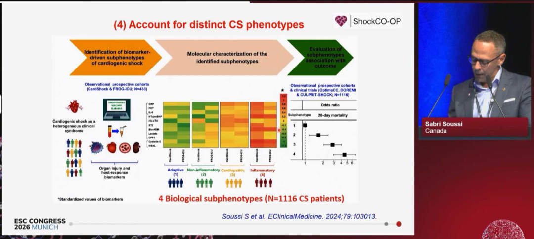 Fig. 5. The four Biological Subtypes Show Significant Differences in 28-day Mortality
Conclusion
Prof. Soussi’s lecture represents a profound reflection on the traditional CS diagnostic model. He emphasized that to shift from “diagnosing established hypoperfusion” to “preventing the occurrence of hypoperfusion,” the key lies in constructing a more sensitive prospective diagnostic system. By integrating ScvO2, LVOT-VTI, and point-of-care testing (POCT) biomarkers, clinicians can precisely identify high-risk patients before organ function is impaired.
From the introduction of the “early shock” concept to the implementation of a multi-dimensional indicator integration system, this marks the definitive departure of cardiogenic shock management from the previous “one-size-fits-all” clinical syndrome model. The field is steadily moving toward a precision medicine era jointly driven by hemodynamics and molecular biology. This evolution of the medical paradigm is not merely a theoretical innovation at the academic level; it will directly reshape future clinical practice, advance clinical trials through more precise subject selection, guide targeted therapeutic directions, and ultimately translate into more individualized management strategies—holding profound landmark significance for improving survival outcomes in patients with cardiogenic shock.

